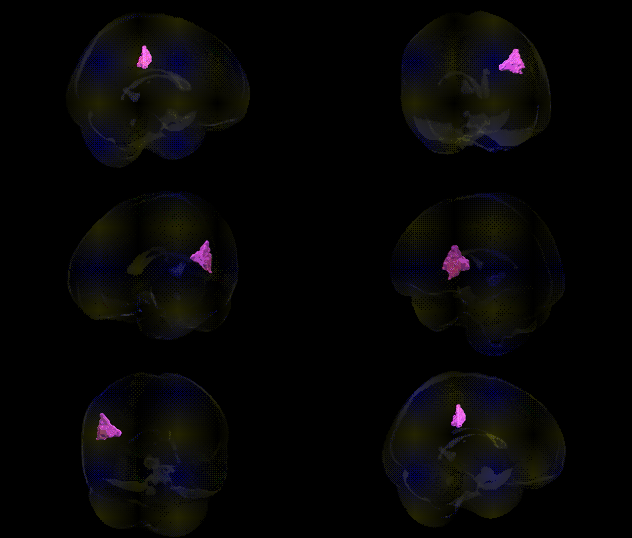
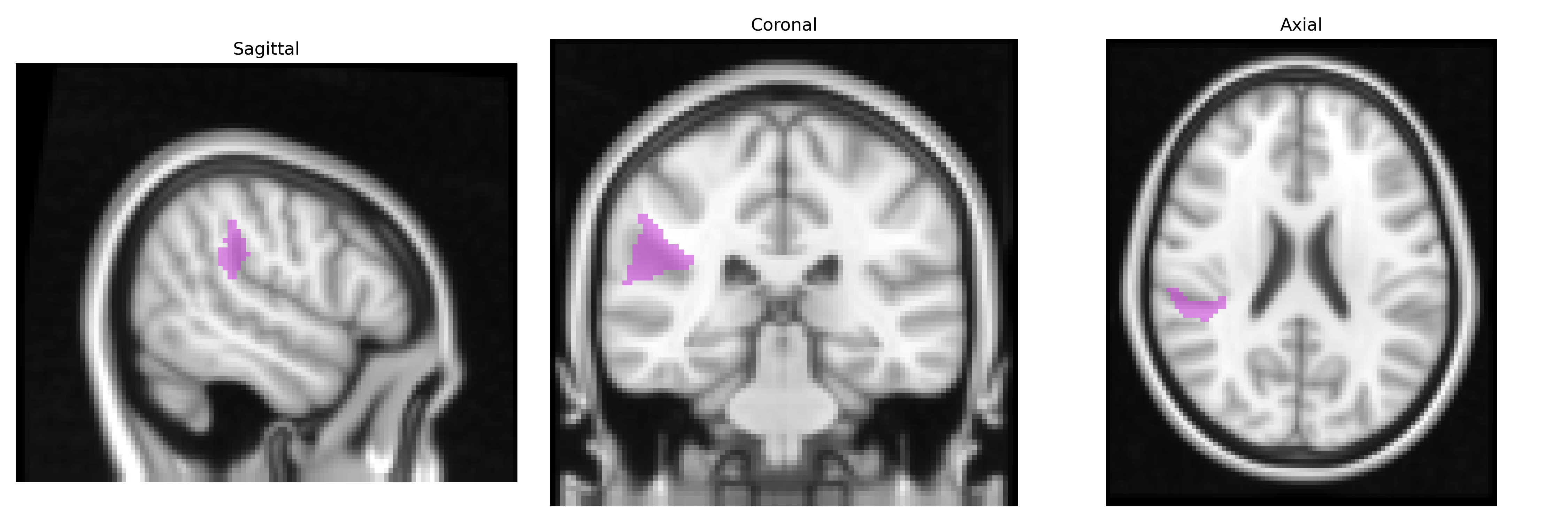
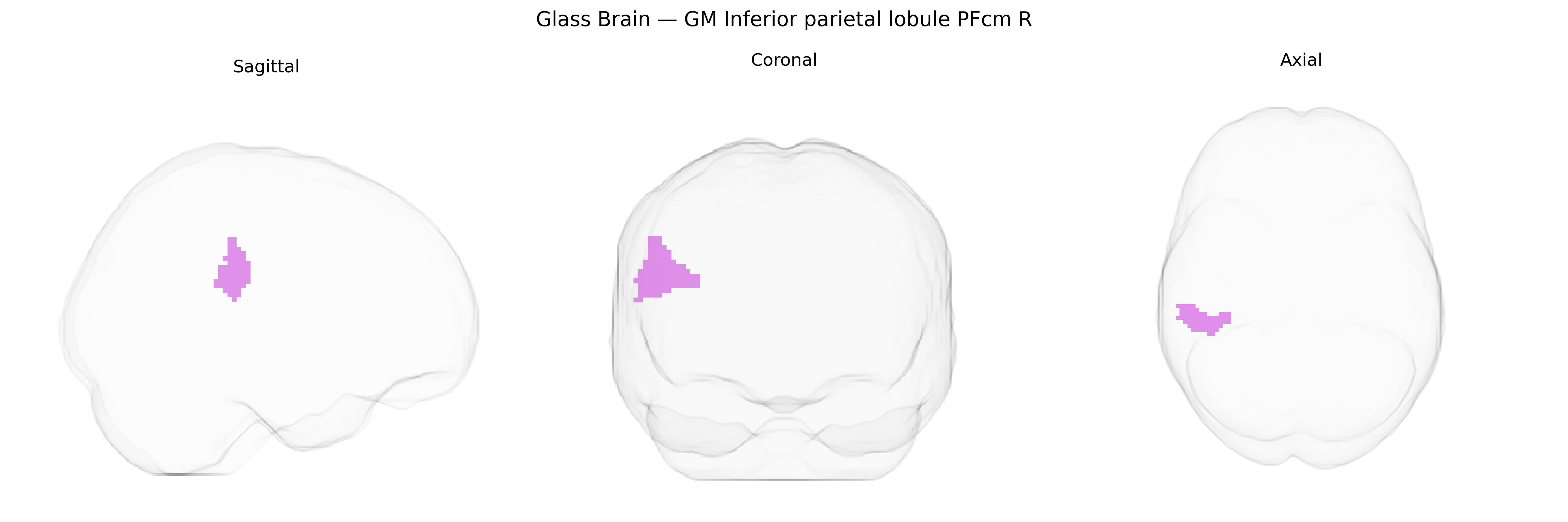
The bilateral GM Inferior parietal lobule PFcm R (Jülich maxprob thr25 2mm atlas) corresponds to a cytoarchitectonically defined subdivision of the inferior parietal lobule (IPL) in the posterior parietal cortex, located near the supramarginal gyrus and bordering other IPL fields such as PF, PFt, and PFop. This region is implicated in multisensory integration, higher-order somatosensory processing, and cognitive functions including attention, action observation, and aspects of social cognition, consistent with the broader role of the IPL within the dorsal stream and temporoparietal networks. PFcm contributes to the transformation of sensory inputs into motor representations, participates in body-related and spatial representation, and is often engaged in tasks involving imitation, tool use, and perspective-taking. There is no direct link for “PFcm,” but it is a subregion of the Inferior parietal lobule.
The bilateral GM Inferior parietal lobule PFcm R (Juelich maxprob thr25 2mm Atlas) lies within the inferior parietal lobule (IPL), a heteromodal association region whose structure and function show substantial heritability and have been repeatedly implicated in imaging–genetics and GWAS-based brain mapping studies. Although no large GWAS to date has isolated PFcm R as a unique genetic target, SNP-based and twin-based studies indicate moderate heritability of cortical thickness, surface area, and gyrification in adjacent IPL subregions, often driven by variants in genes related to synaptic plasticity (e.g., BDNF), axon guidance (e.g., ROBO/Semaphorin signaling), and neuronal development (e.g., NRG1/ERBB pathways). Polygenic risk for schizophrenia, autism spectrum disorder, and major depressive disorder has been associated with structural and functional alterations in nearby inferior parietal territories, including reduced cortical thickness or altered connectivity in right IPL; similarly, risk variants for attention-deficit/hyperactivity disorder and dyslexia show convergent effects on parietal attention and language networks that encompass PFcm-like areas. Large-scale ENIGMA and UK Biobank analyses have linked common variants in genes such as HMGA2, IGF1, and other growth- and neurodevelopment-related loci to global and regional parietal cortical measures that include the PFcm region, and functional imaging genetics has associated parietal activation patterns (particularly during attention, working memory, and social cognition tasks) with dopaminergic and glutamatergic gene variants. Overall, genetic associations for PFcm R are best understood in the context of broader inferior parietal and temporoparietal networks, where common and rare variants influencing neurodevelopment, synaptic signaling, and cortical scaling contribute to susceptibility for neurodevelopmental, psychotic, and mood disorders as well as to interindividual differences in cognitive traits such as language, attention, and numerical processing.
Overview generated by GPT-4o (2026).
Region ID: 30
Hemisphere: bilateral
Atlas: Juelich maxprob thr25 2mm
Full Quality Version: Download MP4
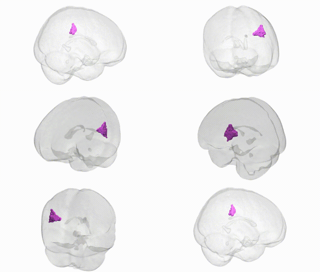
Full Quality Version: Download MP4
Wali Sidiqyar*, Gaurav Rudravaram*, Elyssa M. McMaster, Trent M. Schwartz, Adam M. Saunders, Kurt G. Schilling, Bennett A. Landman "Introducing SPINS: A Shared Public Visualization Library of Neuroanatomical Structures." Medical Imaging with Deep Learning- short paper
This resource is licensed under CC0 1.0 Universal (Public Domain).