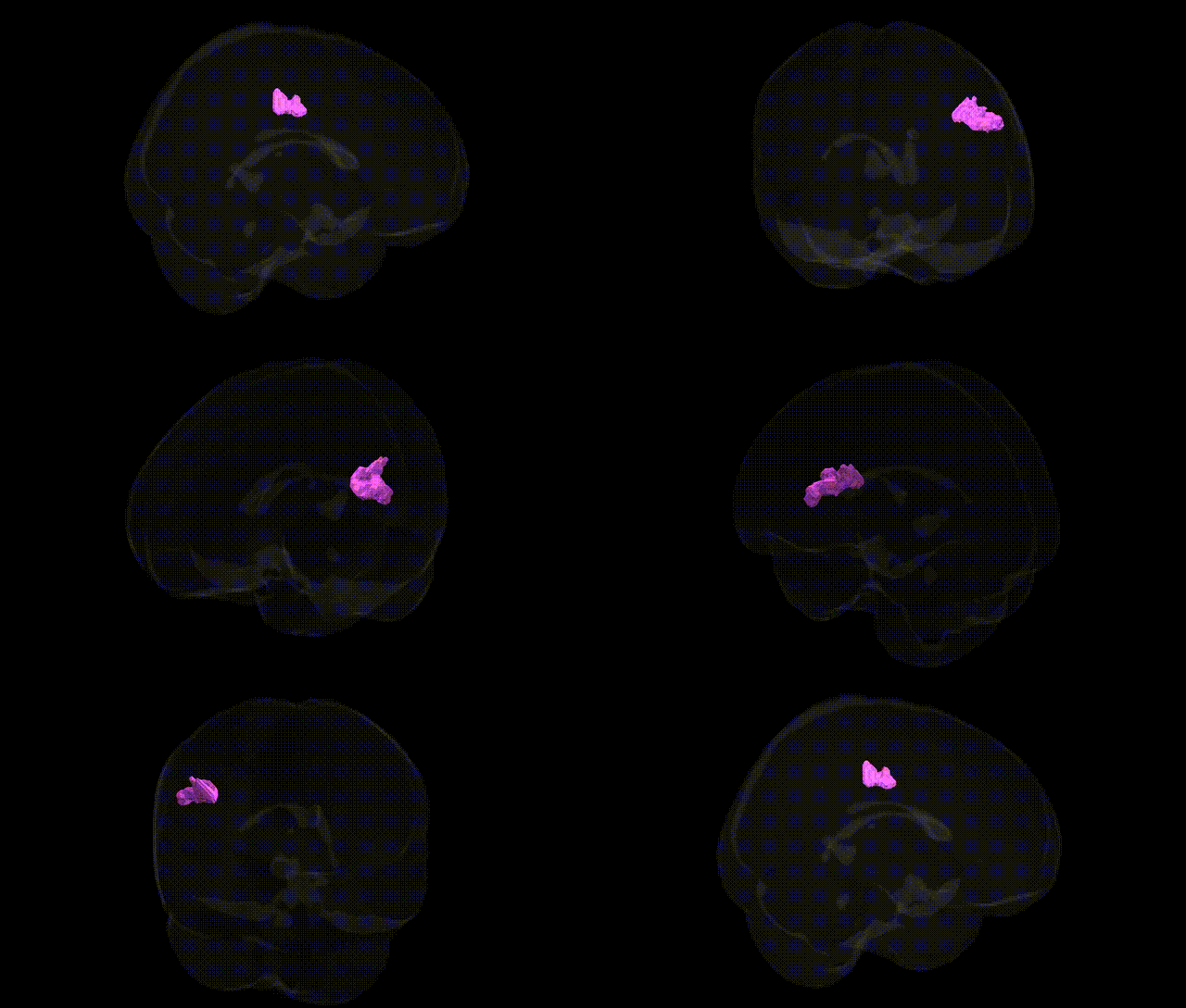
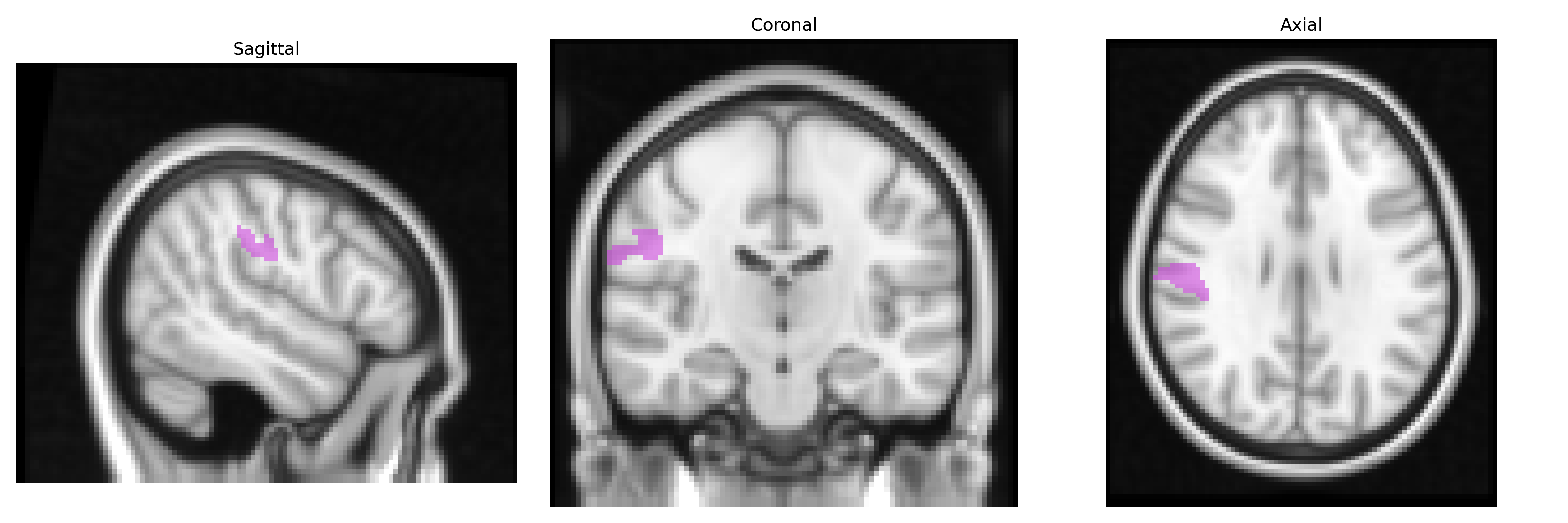
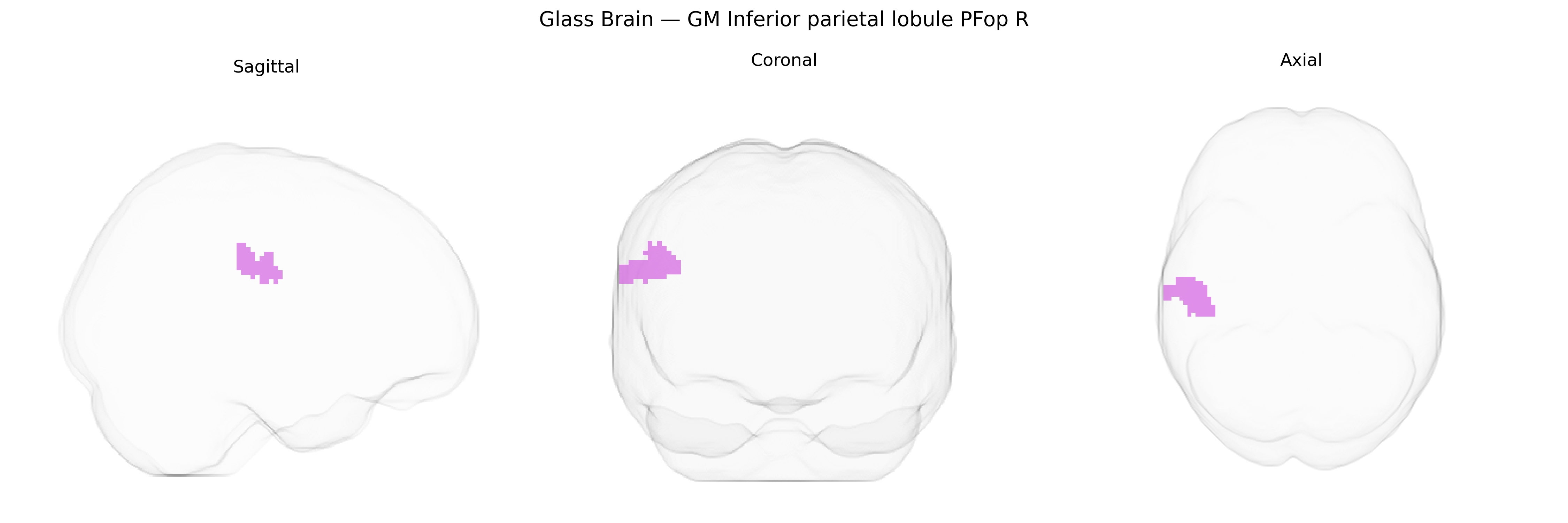
The bilateral GM Inferior parietal lobule PFop R from the Juelich maxprob thr25 2mm Atlas corresponds to a cytoarchitectonic subregion of the inferior parietal lobule (IPL), situated in the posterior part of the supramarginal gyrus along the lateral surface of the parietal lobe, bordering the superior temporal and frontal regions. This area is implicated in multimodal integration of visual, auditory, and somatosensory information and is associated with higher-order cognitive functions such as language processing (including phonological and articulatory components), praxis, attention reorienting, and aspects of social cognition such as action understanding and imitation. Functionally, PFop forms part of a broader fronto-parietal network involved in sensorimotor integration and may participate in the dorsal stream of language and action processing. There is no direct link for PFop, but it is a subregion of the Inferior parietal lobule.
The bilateral inferior parietal lobule, including the PFop subdivision in the right hemisphere as defined in the Juelich maxprob thr25 2mm atlas, has been implicated in multiple genetic and GWAS findings related to higher cognitive function, language, and neuropsychiatric risk, though few studies target PFop specifically at fine-grained cytoarchitectonic resolution. Large-scale imaging genetics consortia (e.g., ENIGMA, UK Biobank) have identified common variants influencing cortical thickness and surface area in parietal association cortex, including loci near genes such as HMGA2, KIAA0586, and variants in neurodevelopmental and synaptic genes that modulate parietal morphology and connectivity. Polygenic scores for general cognitive ability and educational attainment show robust associations with structural and functional measures in inferior parietal regions, which are core hubs for attention, semantic processing, and the default mode/frontoparietal networks. GWAS of language-related traits and reading abilities have highlighted parietal and temporoparietal regions, with genes involved in axon guidance, neuronal migration, and synaptic plasticity (e.g., FOXP2-related networks, DCDC2, CNTNAP2) contributing to variability in activation and structure in inferior parietal cortex during language tasks. Neuropsychiatric GWAS and imaging genetics studies in schizophrenia, autism spectrum disorder, and major depressive disorder have repeatedly reported parietal association cortex, including inferior parietal lobule, as a site of genetically influenced cortical thinning, altered connectivity, or task-related hypo/hyperactivation, linking risk variants in synaptic, glutamatergic, and immune-related genes (such as those in the MHC region and complement pathway) to parietal network dysfunction. In neurodegenerative and age-related conditions, genetic risk for Alzheimer’s disease (e.g., APOE ε4 and other GWAS loci like BIN1, CLU, and PICALM) is associated with accelerated atrophy and metabolic changes in posterior parietal regions that overlap or neighbor PFop, consistent with the role of parietal association cortex in episodic memory and visuospatial functions. Overall, genetic studies converge on the inferior parietal lobule as a multimodal association hub whose structure and function are shaped by polygenic influences on cortical development, synaptic signaling, and network organization, with downstream effects on cognition, language, and vulnerability to psychiatric and neurodegenerative disorders, even though PFop-specific genetic associations remain largely inferred from broader parietal and frontoparietal imaging genetics findings rather than being directly mapped to this single cytoarchitectonic subdivision.
Overview generated by GPT-4o (2026).
Region ID: 34
Hemisphere: bilateral
Atlas: Juelich maxprob thr25 2mm
Full Quality Version: Download MP4
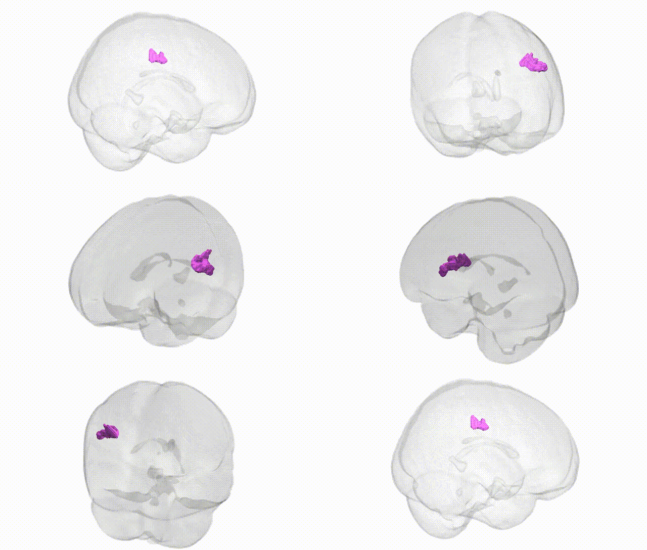
Full Quality Version: Download MP4
Wali Sidiqyar*, Gaurav Rudravaram*, Elyssa M. McMaster, Trent M. Schwartz, Adam M. Saunders, Kurt G. Schilling, Bennett A. Landman "Introducing SPINS: A Shared Public Visualization Library of Neuroanatomical Structures." Medical Imaging with Deep Learning- short paper
This resource is licensed under CC0 1.0 Universal (Public Domain).