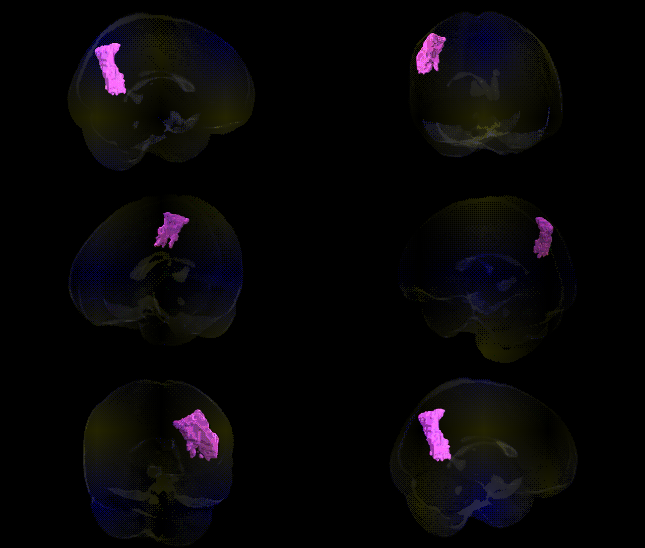
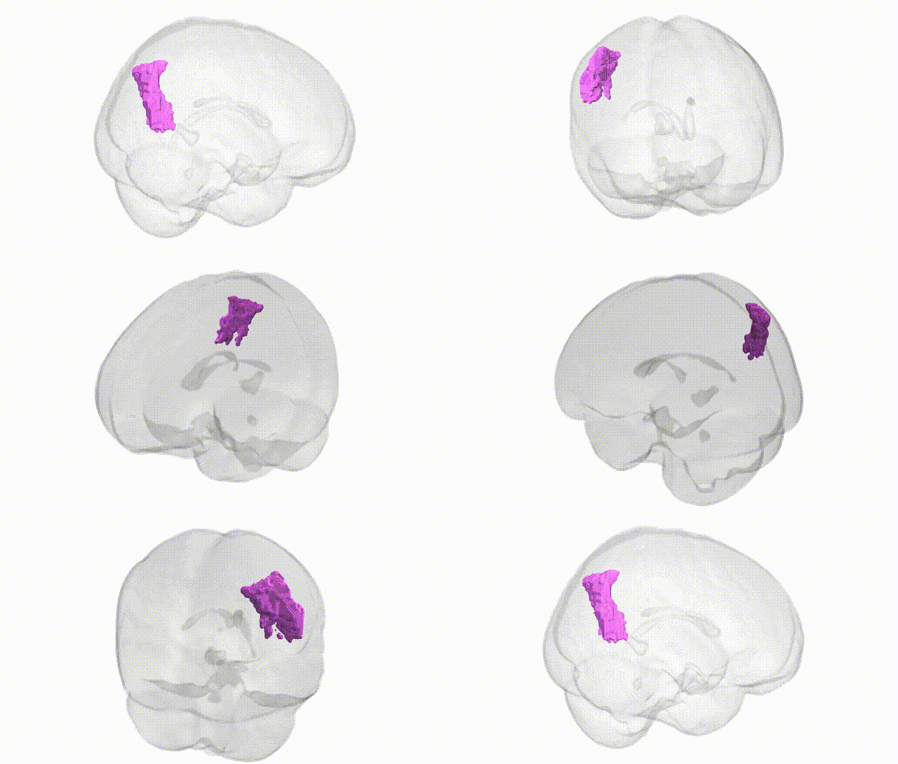
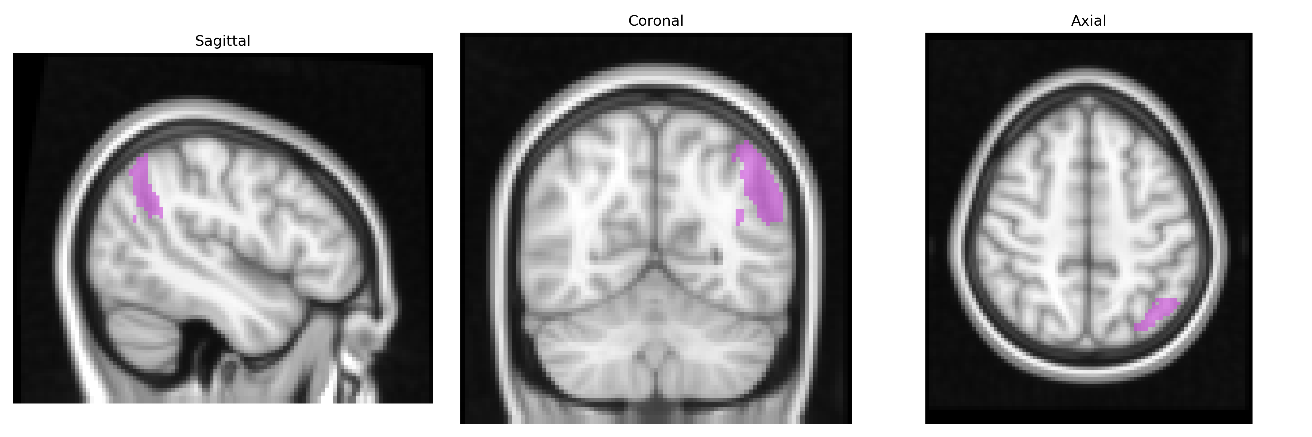
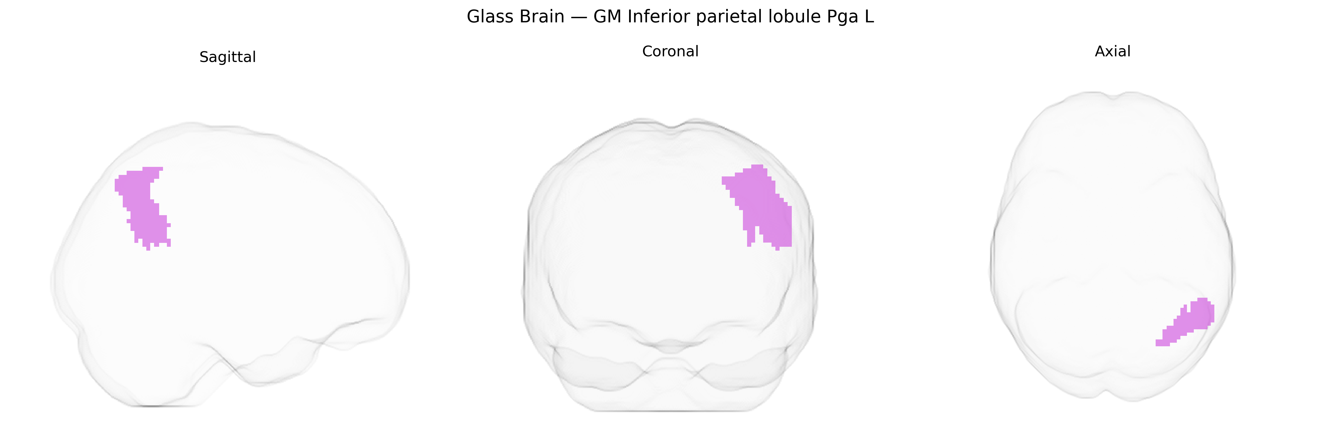
The bilateral GM Inferior parietal lobule PGa L, as defined in the Juelich maxprob thr25 2mm Atlas, corresponds to the left-hemispheric portion of cytoarchitectonic area PGa within the inferior parietal lobule, a multimodal association region implicated in higher-order cognitive integration. This area is located in the posterior parietal cortex, bordering the supramarginal and angular gyri, and is characterized by dense connections with frontal, temporal, and occipital cortices, supporting functions such as language-related processing, semantic integration, spatial attention, and aspects of social cognition. Neuronal populations in PGa show responsiveness to convergent visual, auditory, and somatosensory inputs, enabling cross-modal integration and contribution to body representation and action understanding. There is no direct Wikipedia article for PGa; a related structure is the Inferior parietal lobule.
The bilateral GM Inferior parietal lobule PGa (left hemisphere) in the Juelich maxprob thr25 2 mm atlas corresponds to a higher-order association area implicated in language, attention, and social cognition, and genetic associations identified for inferior parietal regions more broadly converge on neurodevelopmental and psychiatric traits rather than on this precise subfield alone. GWAS and imaging-genetics studies have shown that common variants influencing cortical surface area and thickness in inferior parietal regions include loci near genes such as HMGA2, RSPO3, and factors in Wnt and neurodevelopmental pathways, and that polygenic scores for educational attainment, intelligence, and general cognitive ability are associated with structural variation in inferior parietal cortex. Variants in genes involved in synaptic plasticity and glutamatergic signaling (for example, CACNA1C and GRIN family genes in large-scale psychiatric GWAS) have been linked to structural and functional alterations in inferior parietal lobule in schizophrenia, bipolar disorder, and major depression, while ASD- and ADHD-related polygenic risk has been associated with altered morphology and connectivity in parietal association cortices, including inferior parietal regions. Additional imaging-genetics work links inferior parietal structure and function to Alzheimer’s disease risk loci (such as APOE and CLU), consistent with this region’s involvement in default mode and memory-related networks, and to variants associated with handedness and language lateralization, although most findings are at the level of broader parietal or temporoparietal association cortex rather than specifically the Juelich-defined PGa subregion.
Overview generated by GPT-4o (2026).
Region ID: 37
Hemisphere: bilateral
Atlas: Juelich maxprob thr25 2mm
Full Quality Version: Download MP4
Full Quality Version: Download MP4
Wali Sidiqyar*, Gaurav Rudravaram*, Elyssa M. McMaster, Trent M. Schwartz, Adam M. Saunders, Kurt G. Schilling, Bennett A. Landman "Introducing SPINS: A Shared Public Visualization Library of Neuroanatomical Structures." Medical Imaging with Deep Learning- short paper
This resource is licensed under CC0 1.0 Universal (Public Domain).