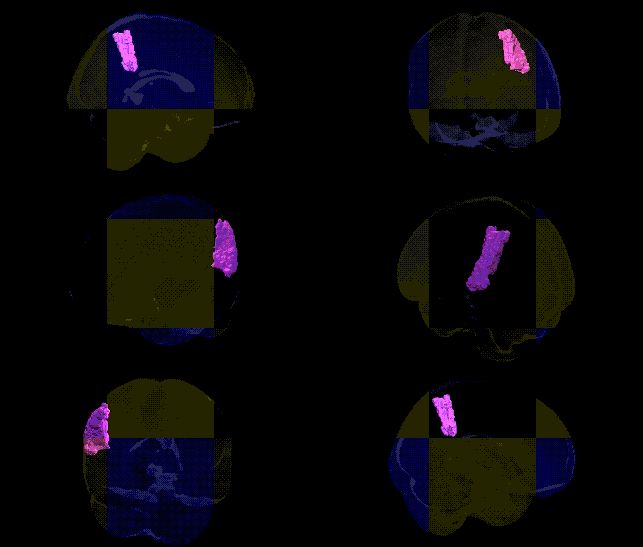
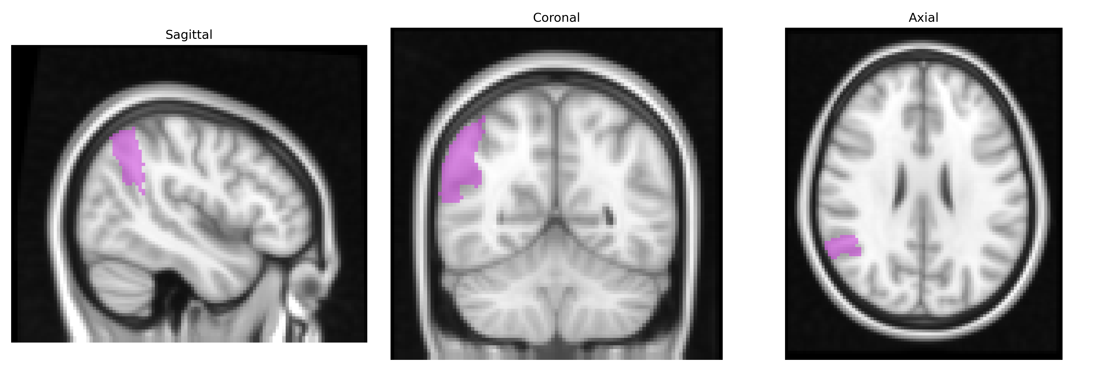
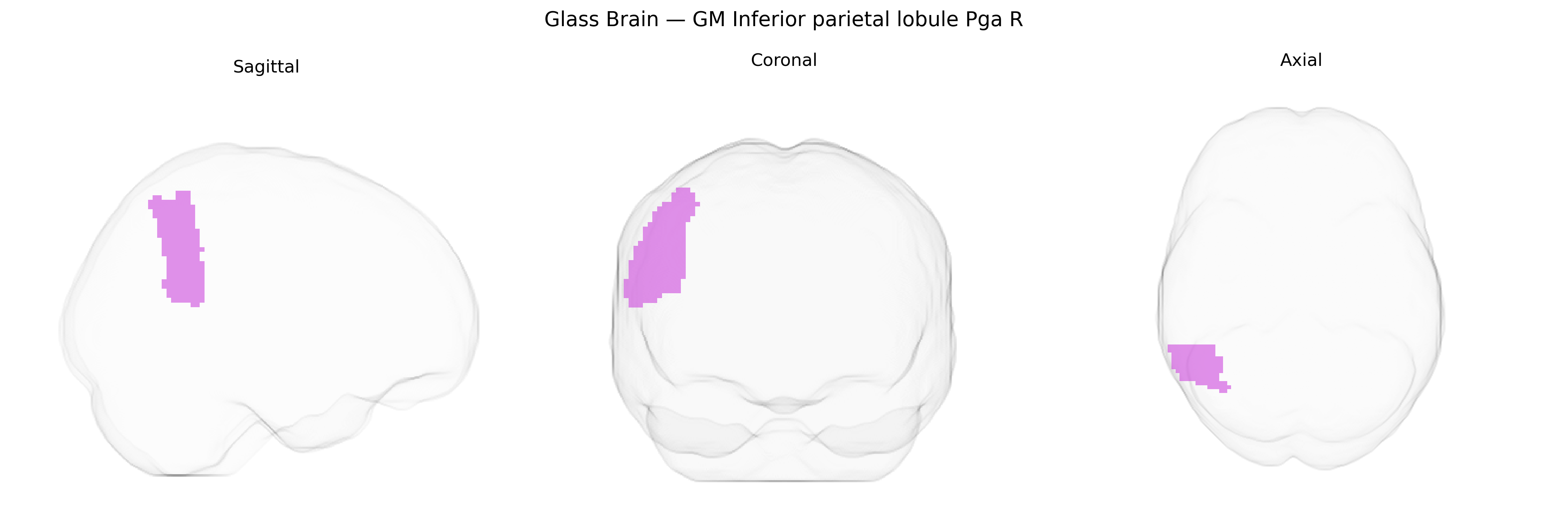
The bilateral GM Inferior parietal lobule PGa R, defined in the Juelich maxprob thr25 2 mm atlas, corresponds to the right-hemispheric granular subdivision A (PGa) of the inferior parietal lobule, a higher-order association area located at the caudal end of the supramarginal and angular gyri. Cytoarchitectonically, PGa is part of the inferior parietal cortex that integrates multimodal sensory input, including visual, auditory, and somatosensory information, and is implicated in functions such as spatial attention, language processing, semantic integration, and aspects of social cognition and theory of mind. Connectivity studies indicate strong reciprocal links with frontal association cortices, temporal lobe language regions, and occipital visual areas, supporting a role in the dorsal attention and default mode networks. There is no direct Wikipedia article for PGa; a related structure is the Inferior parietal lobule.
The bilateral GM Inferior parietal lobule PGa (right) in the Juelich maxprob thr25 2 mm atlas corresponds to a higher-order multimodal association area frequently implicated in genetic studies of cortical morphology, cognition, and neuropsychiatric risk. GWAS of brain structure (e.g., ENIGMA, UK Biobank) have repeatedly identified common variants near genes involved in neurodevelopment, synaptic function, and axon guidance—such as MIR137, DCC, CELSR3, and POU3F2—associated with surface area, thickness, or volume in inferior parietal or adjacent temporo‑parietal regions, with some loci showing pleiotropy across multiple association cortices. Polygenic scores for educational attainment and general cognitive ability show positive associations with increased cortical surface area or thickness in inferior parietal regions, aligning with this area’s role in language, number processing, and attentional control. Conversely, schizophrenia-, bipolar disorder-, and major depression–related polygenic risk scores have been linked to altered morphology and functional connectivity in inferior parietal cortex, and specific risk loci (e.g., CACNA1C, GRIN2A, complement pathway genes) have shown associations with temporo‑parietal structural and functional metrics in case–control and imaging–genetics studies. Additional genetic correlations connect inferior parietal variation with traits such as autism spectrum disorder (social cognition networks), ADHD (attention networks), Alzheimer’s disease and amyloid/tau pathology (parietal atrophy patterns, especially in APOE ε4 carriers), and chronic pain or somatosensory traits, though these associations often involve broader parietal or default mode network regions rather than the PGa subfield alone.
Overview generated by GPT-4o (2026).
Region ID: 38
Hemisphere: bilateral
Atlas: Juelich maxprob thr25 2mm
Full Quality Version: Download MP4
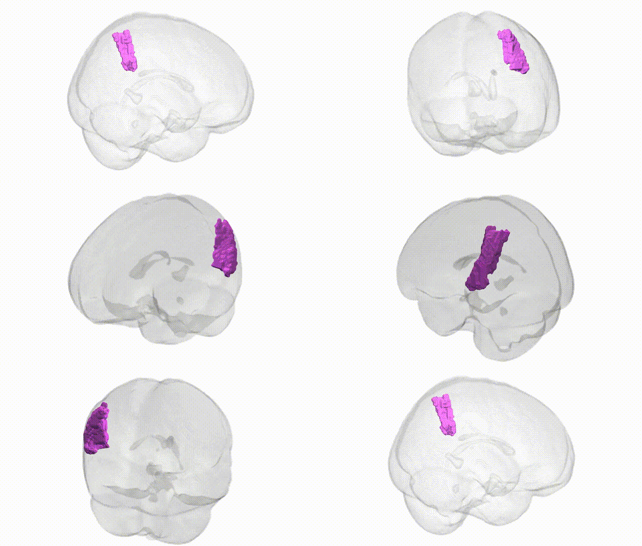
Full Quality Version: Download MP4
Wali Sidiqyar*, Gaurav Rudravaram*, Elyssa M. McMaster, Trent M. Schwartz, Adam M. Saunders, Kurt G. Schilling, Bennett A. Landman "Introducing SPINS: A Shared Public Visualization Library of Neuroanatomical Structures." Medical Imaging with Deep Learning- short paper
This resource is licensed under CC0 1.0 Universal (Public Domain).