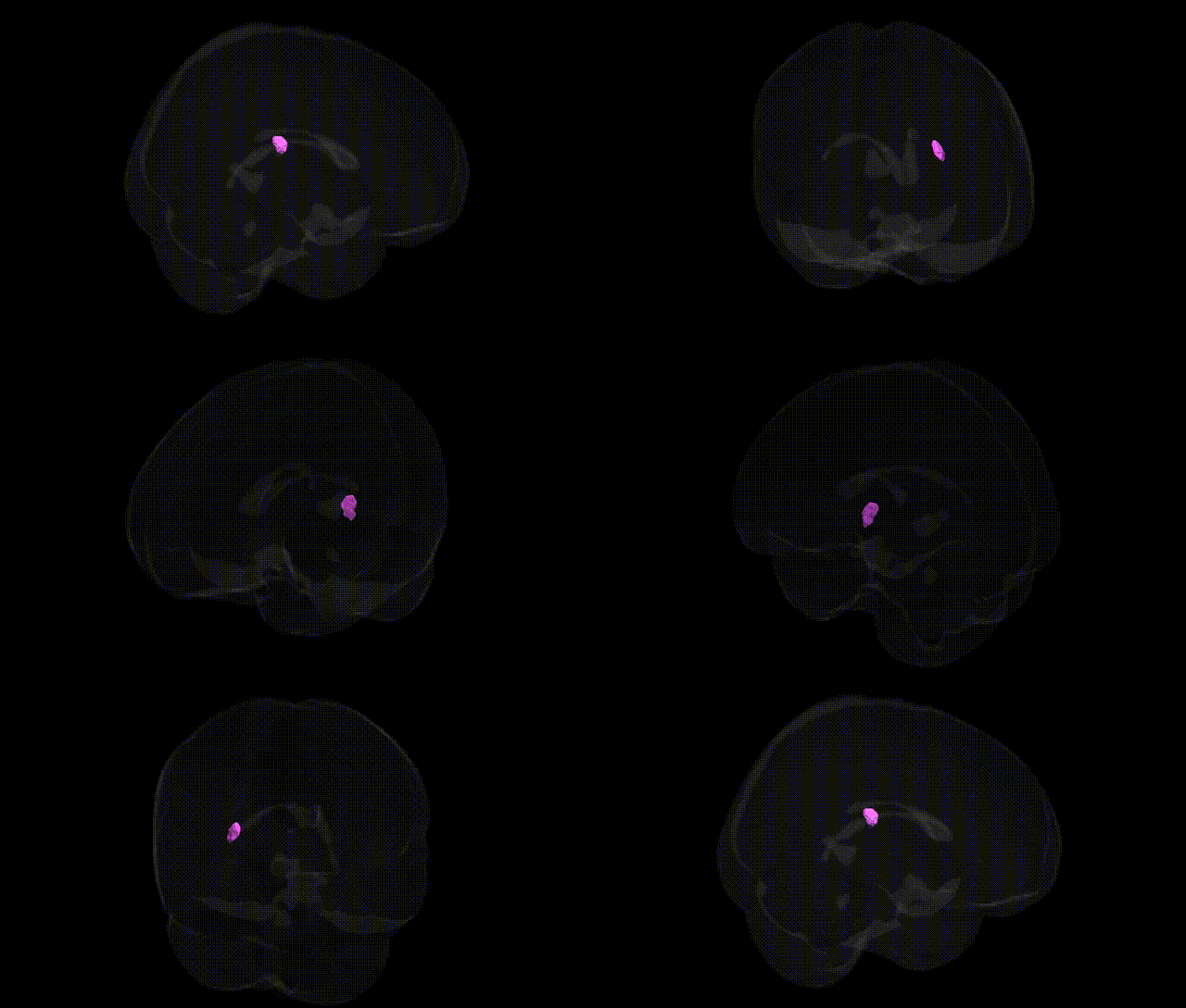
The bilateral GM Insula Ig1 R region in the Juelich maxprob thr25 2 mm atlas corresponds to the granular insular cortex, a primary sensory subdivision of the insula located deep within the lateral sulcus beneath the frontal, parietal, and temporal opercula. Cytoarchitectonically, Ig1 is characterized by a well-developed granular layer IV and distinct laminar organization, distinguishing it from agranular and dysgranular insular areas. Functionally, this granular insular territory is implicated in multimodal sensory integration, including viscerosensory and somatosensory processing, and contributes to interoception and the representation of bodily states that support homeostatic regulation and affective experience. As part of the broader insular cortex, Ig1 participates in salience detection and the coordination of autonomic responses via extensive connections with limbic, prefrontal, and somatosensory regions. There is no direct Wikipedia article for “Insula Ig1”; a closely related structure is the Insular cortex.
The bilateral GM Insula Ig1 R region in the Juelich maxprob thr25 2mm atlas corresponds to anterior–mid insular cortex, a multimodal integration hub repeatedly implicated in genetically informed studies of brain structure and function. Large-scale imaging genetics GWAS (e.g., ENIGMA, UK Biobank) have shown that insular cortical thickness and surface area are heritable and associated with variants in genes involved in neuronal development, synaptic signaling, and neuroplasticity, including loci near genes such as DLG2, TESC, and others implicated in cortical morphometry. Insular morphology and connectivity have been linked to polygenic risk for psychiatric disorders, including major depressive disorder, schizophrenia, bipolar disorder, and anxiety, where risk alleles often predict altered insular volume or activity, especially in salience and interoceptive networks. GWAS of smoking behavior, alcohol use, and obesity-related traits have found that polygenic scores for these behaviors correlate with insular structure and activation, consistent with the region’s role in addiction, craving, and taste/interoception; some loci (e.g., in CHRNA5–CHRNA3–CHRNB4 for smoking) show downstream associations with insula-related circuitry. Insula Ig1–like territories (anterior/mid insula) also appear in imaging genetics studies of pain sensitivity, empathy and social cognition, and autonomic regulation, where genetic variation in serotonin, dopamine, and opioid system genes modulates insular responses. While specific SNP-level associations uniquely and exclusively tied to the Ig1 R subregion are not well resolved, convergent GWAS and polygenic risk studies consistently implicate this insular territory in the genetically influenced architecture of affect regulation, salience processing, addiction, and multiple neuropsychiatric disorders.
Overview generated by GPT-4o (2026).
Region ID: 119
Hemisphere: bilateral
Atlas: Juelich maxprob thr25 2mm
Full Quality Version: Download MP4
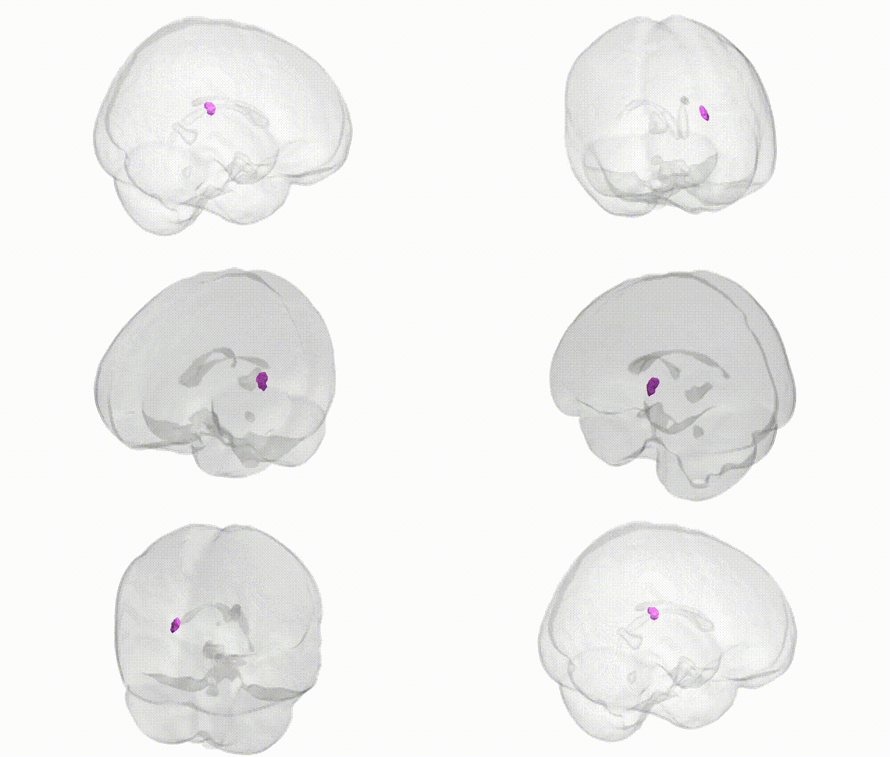
Full Quality Version: Download MP4
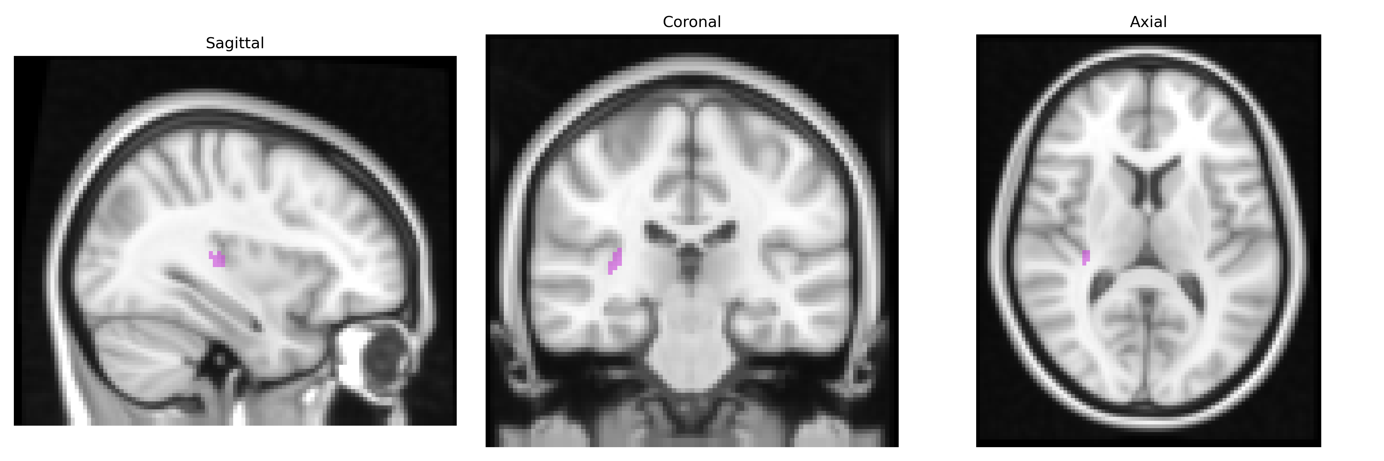
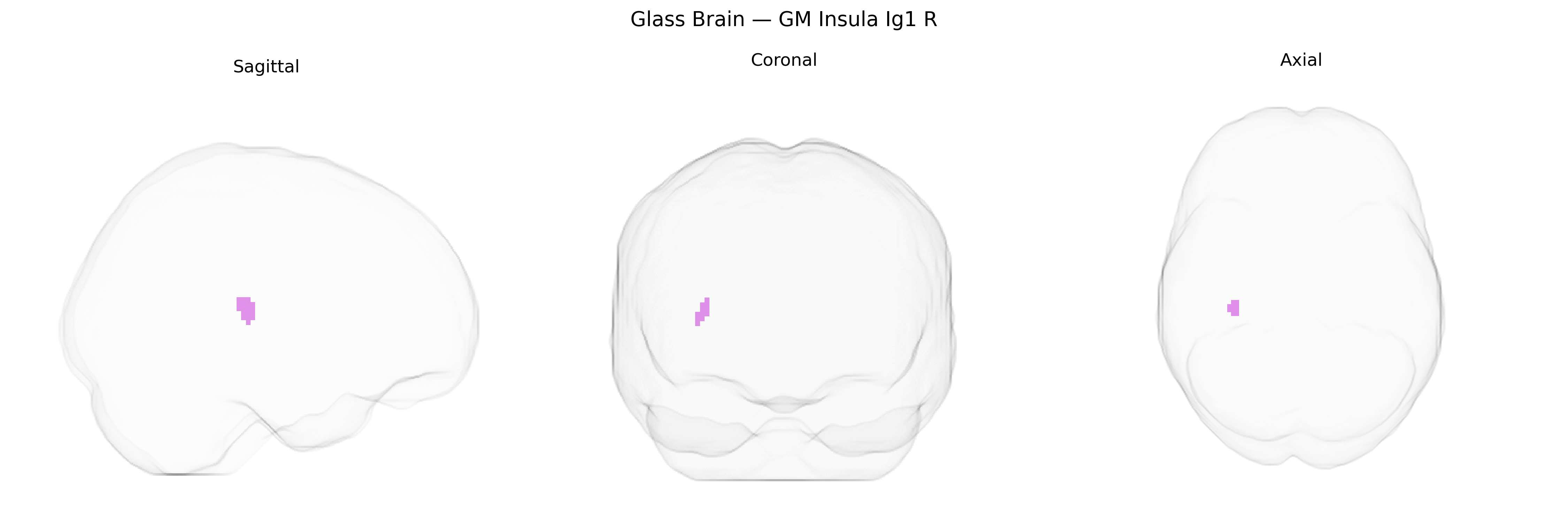
Wali Sidiqyar*, Gaurav Rudravaram*, Elyssa M. McMaster, Trent M. Schwartz, Adam M. Saunders, Kurt G. Schilling, Bennett A. Landman "Introducing SPINS: A Shared Public Visualization Library of Neuroanatomical Structures." Medical Imaging with Deep Learning- short paper
This resource is licensed under CC0 1.0 Universal (Public Domain).