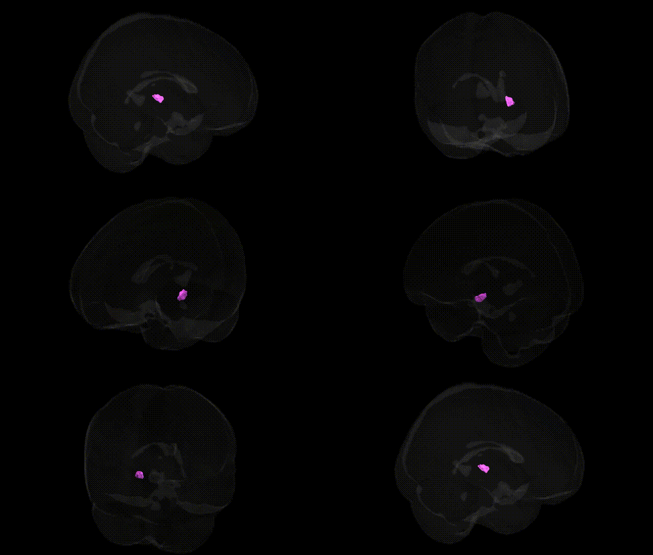
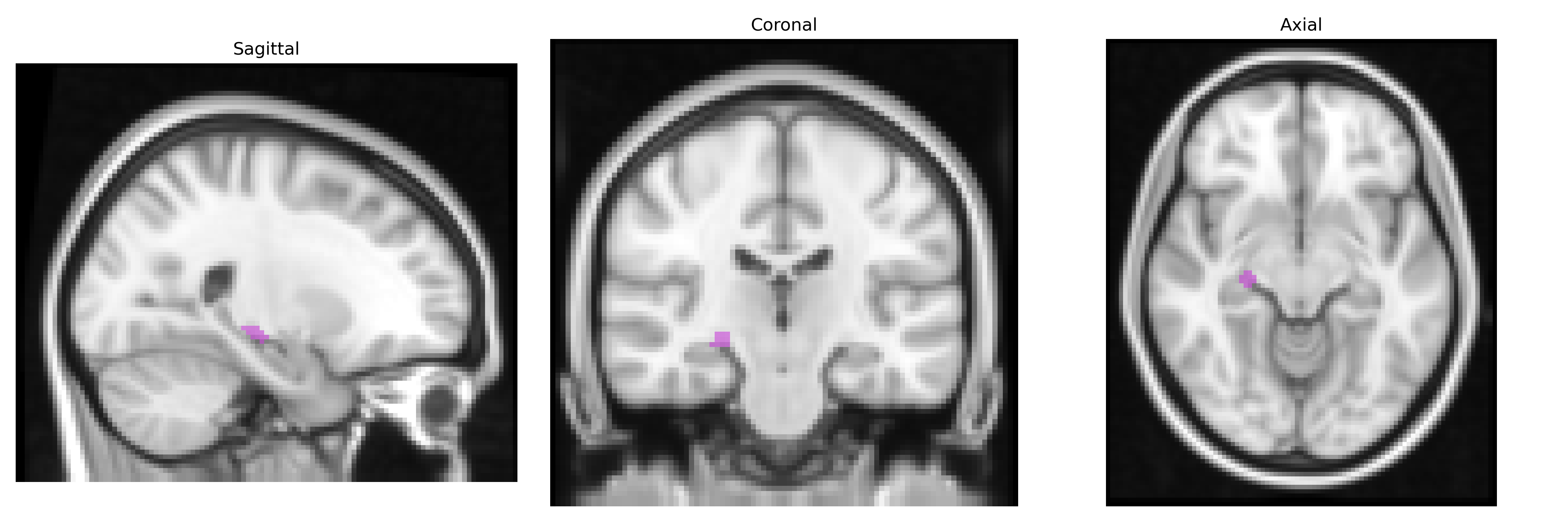
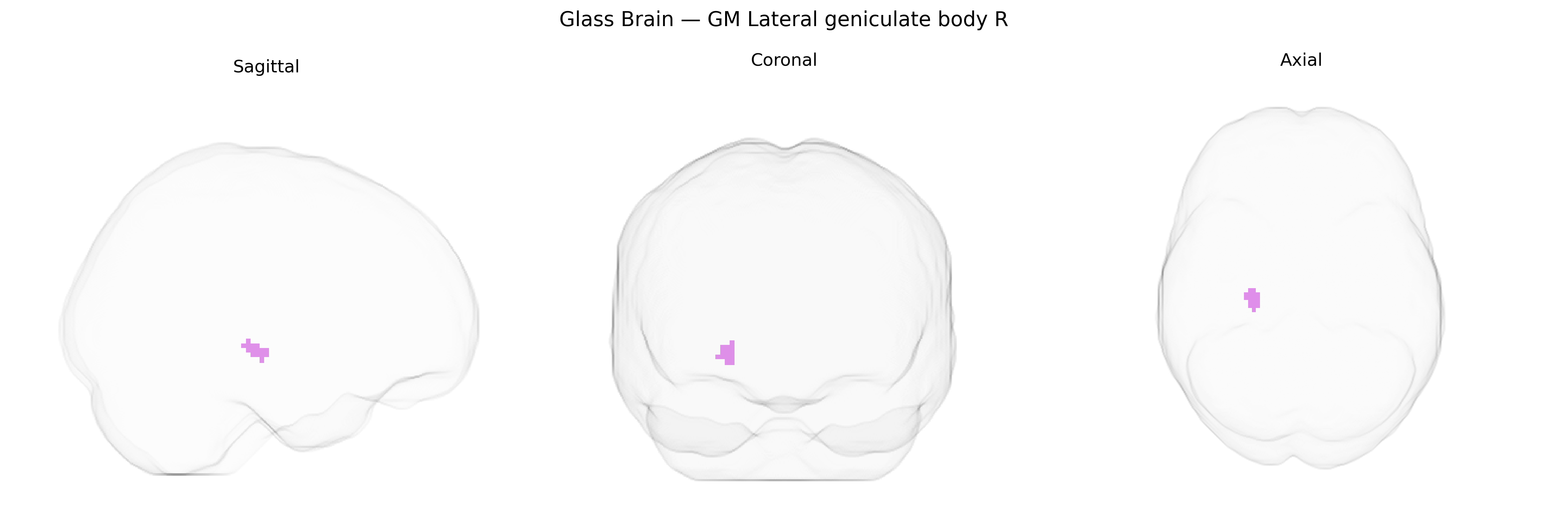
The bilateral GM Lateral geniculate body R, as defined in the Juelich maxprob thr25 2 mm Atlas, corresponds to the right lateral geniculate nucleus (LGN) of the thalamus, a laminated relay structure in the dorsal thalamus that receives direct retinal input from the optic tract and projects via the optic radiation to the primary visual cortex (V1, Brodmann area 17). It is organized into distinct magnocellular, parvocellular, and koniocellular layers that process complementary aspects of visual information, including luminance contrast, motion, spatial resolution, and color, with inputs segregated by eye (ocular dominance) and by type of retinal ganglion cell. Functionally, this nucleus plays a central role in the early stages of the visual pathway, modulating and gating retinal signals under the influence of cortical feedback and other subcortical inputs, and is essential for normal conscious visual perception. Lateral geniculate nucleus
The bilateral gray matter lateral geniculate body (LGB) in the Juelich maxprob thr25 2mm atlas corresponds to the thalamic relay nucleus for visual information, and genetic associations involving this region arise mainly from imaging-genetics and neuro-ophthalmologic studies rather than LGB-specific GWAS. Variants influencing visual pathway structure and function—such as those in genes related to myelination (e.g., MAG, MBP), axonal guidance (e.g., ROBOs, EPHs), and synaptic transmission (e.g., GRIN family)—have been implicated in volumetric and microstructural differences along the optic radiations and thalamic visual nuclei, including the LGN/LGB, in large neuroimaging cohorts like UK Biobank, though typically reported at the level of broader thalamic or visual system measures rather than the LGB alone. In disease-focused genetics, loci associated with glaucoma (e.g., SIX6, CDKN2B-AS1), optic neuritis in multiple sclerosis (HLA region and immune-related genes), and mitochondrial disorders affecting the optic nerve (e.g., MT-ND genes in Leber hereditary optic neuropathy) have been linked to secondary structural and functional alterations in the LGB/visual thalamus observed with MRI and diffusion imaging. Neurodevelopmental and neuropsychiatric GWAS (e.g., for schizophrenia, bipolar disorder, autism) occasionally show downstream involvement of thalamic sensory nuclei, including the LGB, via polygenic influences on thalamocortical connectivity, but these effects are diffuse and not specific to this nucleus. Overall, current genetic evidence connects the Juelich-defined LGB primarily through genes and variants that affect the integrity of the visual pathway, thalamic structure, and neuroimmune or neurodevelopmental processes, rather than through GWAS hits targeted explicitly to the lateral geniculate body as an isolated region.
Overview generated by GPT-4o (2026).
Region ID: 103
Hemisphere: bilateral
Atlas: Juelich maxprob thr25 2mm
Full Quality Version: Download MP4
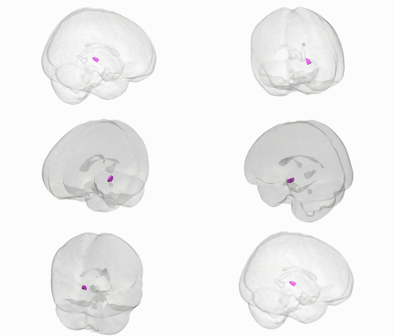
Full Quality Version: Download MP4
Wali Sidiqyar*, Gaurav Rudravaram*, Elyssa M. McMaster, Trent M. Schwartz, Adam M. Saunders, Kurt G. Schilling, Bennett A. Landman "Introducing SPINS: A Shared Public Visualization Library of Neuroanatomical Structures." Medical Imaging with Deep Learning- short paper
This resource is licensed under CC0 1.0 Universal (Public Domain).