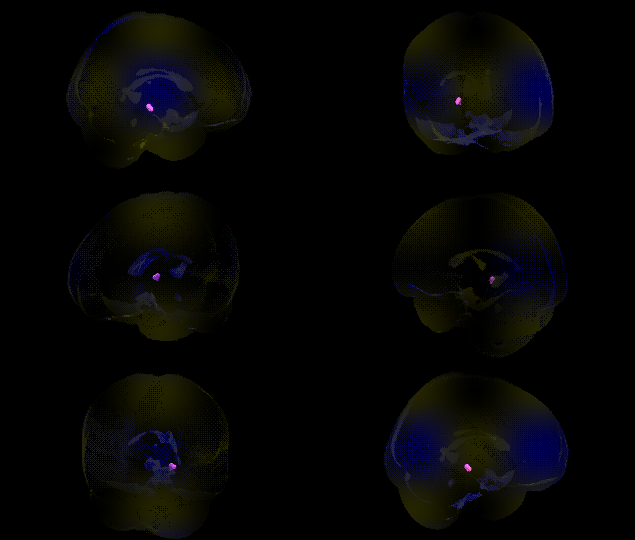
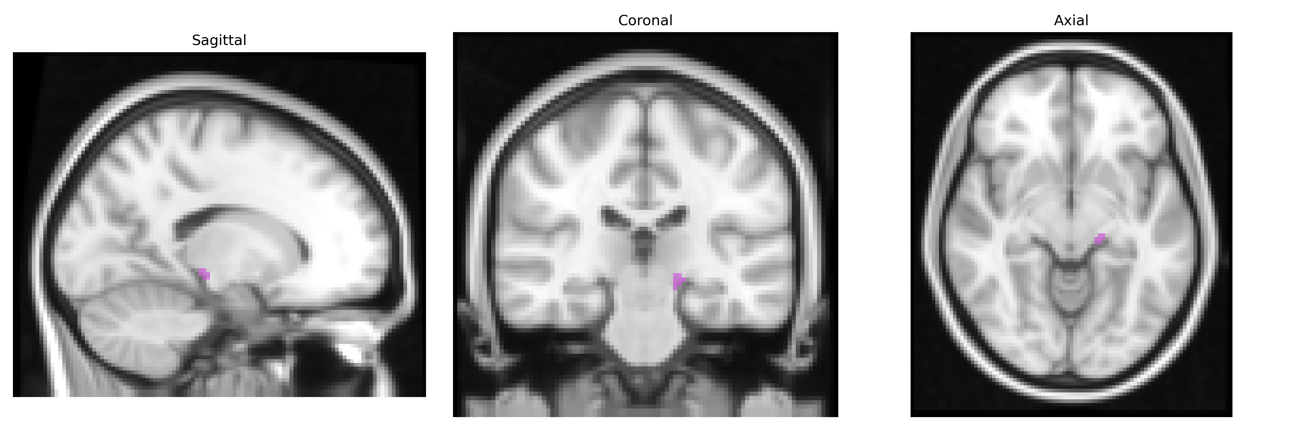
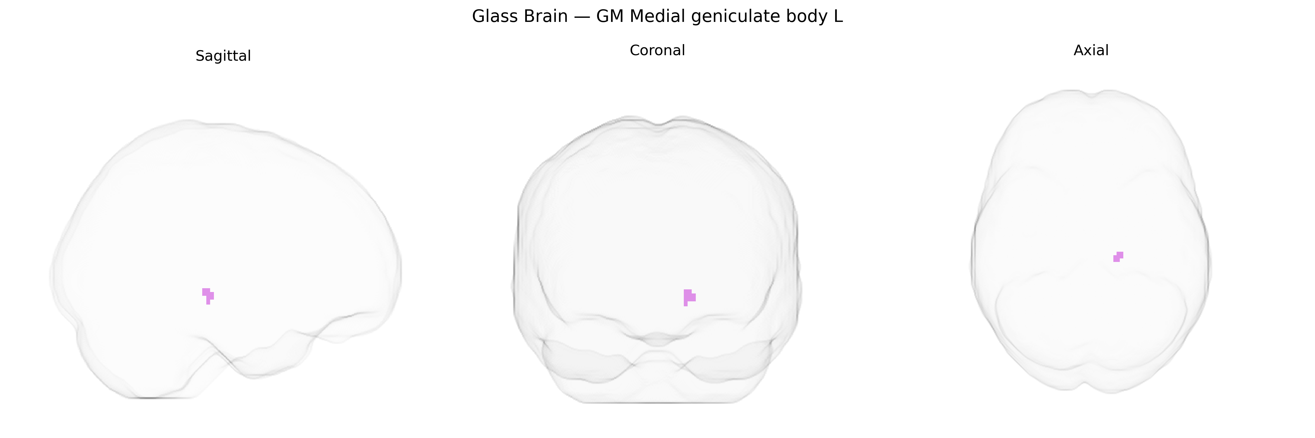
The bilateral GM Medial geniculate body L, as defined in the Juelich maxprob thr25 2 mm atlas, corresponds to the gray matter of the left medial geniculate nucleus (MGN) of the thalamus, a key relay station in the auditory pathway. Located in the posterior thalamus, the medial geniculate body receives ascending input from the inferior colliculus via the brachium of the inferior colliculus and projects primarily to the primary and secondary auditory cortices in the superior temporal lobe, thus mediating the transfer and refinement of auditory information related to sound frequency, intensity, and temporal patterns. It participates in both lemniscal (primary) and non-lemniscal (association) auditory pathways, contributing to functions such as sound localization, auditory attention, and the integration of auditory signals with other sensory and cognitive processes. The region is cytoarchitectonically and functionally heterogeneous, typically subdivided into ventral, dorsal, and medial divisions, each with distinct connectivity and roles in auditory processing. Medial geniculate body
The medial geniculate body (MGB), the thalamic auditory relay corresponding to the GM Medial geniculate body L in the Juelich maxprob atlas, has limited direct region-specific genetic association data, but several convergent findings implicate it via broader auditory, language, and neuropsychiatric pathways. GWAS and imaging-genetics studies of subcortical volumes (e.g., ENIGMA consortium) have identified common variants influencing thalamic and auditory-related structures, though specific MGB-focused hits are rare and typically embedded within larger thalamic or auditory cortex measures. Genetic variants associated with schizophrenia, bipolar disorder, and major depression in large GWAS (e.g., loci in CACNA1C, GRIN2A, and other glutamatergic and calcium-channel genes) have been functionally linked to thalamocortical circuitry, with structural and functional MRI work showing altered MGB responses and connectivity in psychosis and auditory hallucinations, suggesting an indirect genetic–circuit relationship. In autism spectrum disorder and dyslexia, risk genes involved in synaptic development and axon guidance (such as CNTNAP2, FOXP2-related pathways, and others identified in neurodevelopmental GWAS) have been associated with atypical auditory processing and speech perception, where functional imaging frequently implicates MGB and its projections to auditory cortex. Tinnitus and sensorineural hearing loss GWAS highlight genes affecting cochlear function and central auditory plasticity (e.g., KCNQ4 and several noncoding regulatory loci), with thalamic nuclei including MGB implicated in maladaptive gain and phantom sound perception in imaging studies. Overall, while few GWAS report associations specifically labeled to the MGB, genetic influences on thalamocortical and auditory networks—spanning neurodevelopmental, psychiatric, and sensory traits—likely converge on this structure as a key relay in genetically modulated auditory and language-related phenotypes.
Overview generated by GPT-4o (2026).
Region ID: 107
Hemisphere: bilateral
Atlas: Juelich maxprob thr25 2mm
Full Quality Version: Download MP4
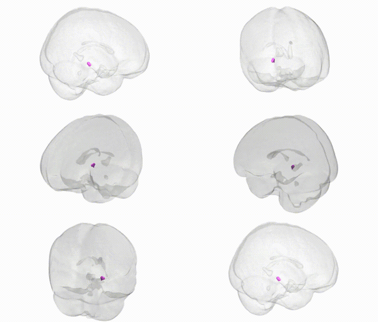
Full Quality Version: Download MP4
Wali Sidiqyar*, Gaurav Rudravaram*, Elyssa M. McMaster, Trent M. Schwartz, Adam M. Saunders, Kurt G. Schilling, Bennett A. Landman "Introducing SPINS: A Shared Public Visualization Library of Neuroanatomical Structures." Medical Imaging with Deep Learning- short paper
This resource is licensed under CC0 1.0 Universal (Public Domain).