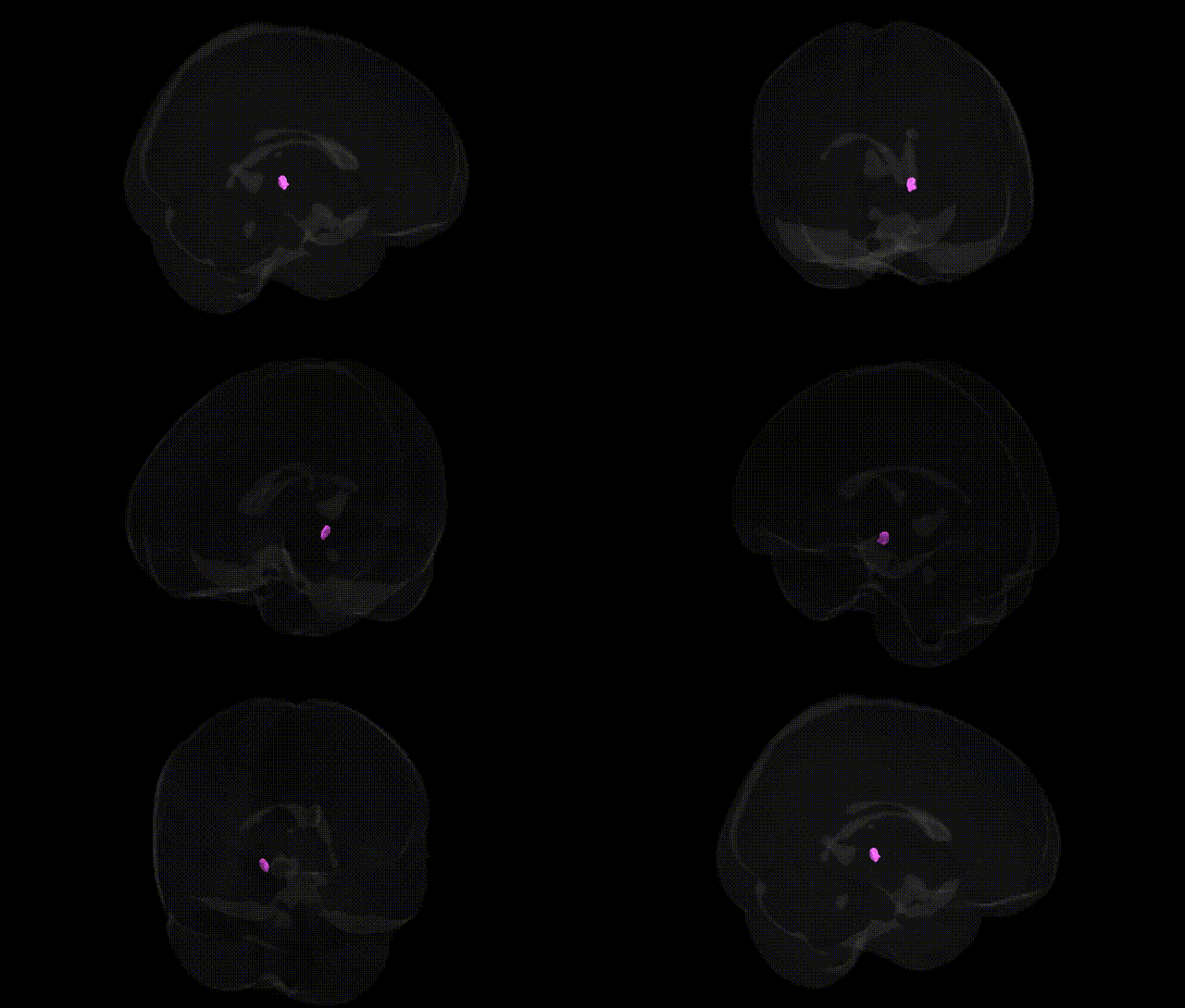
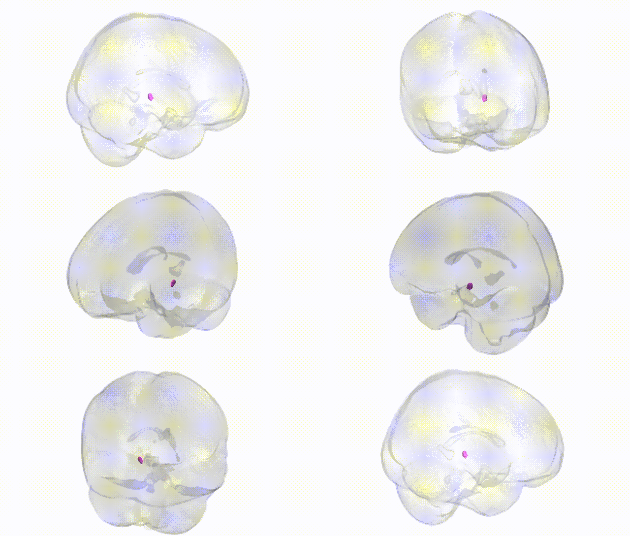
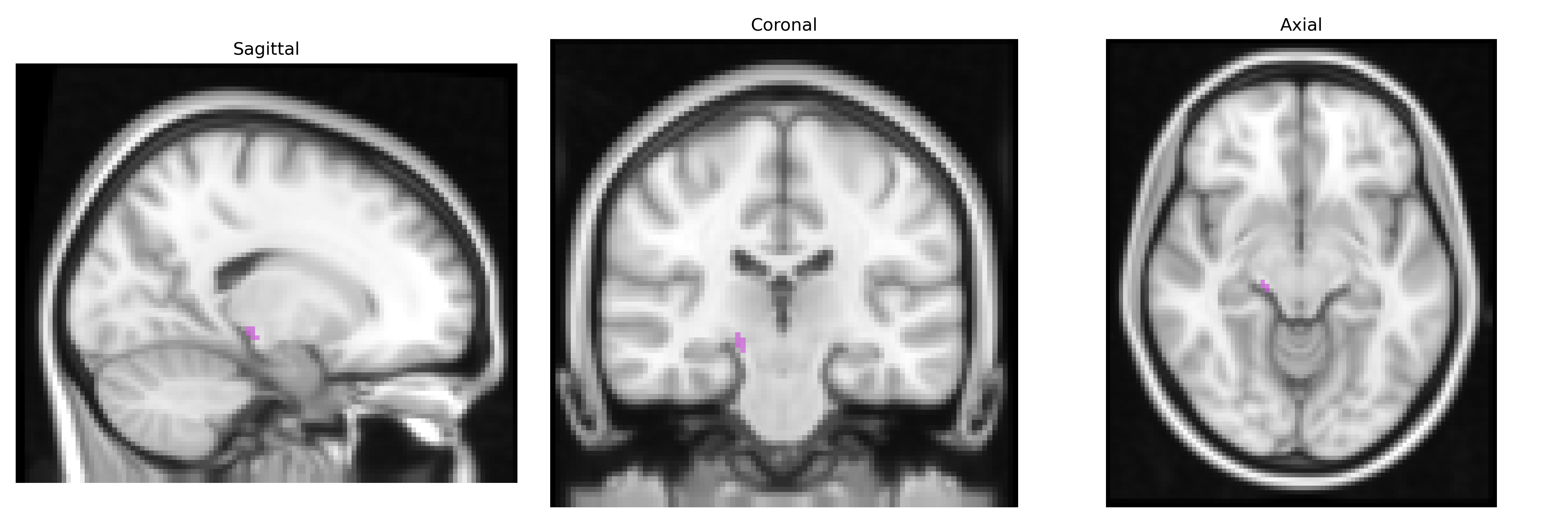
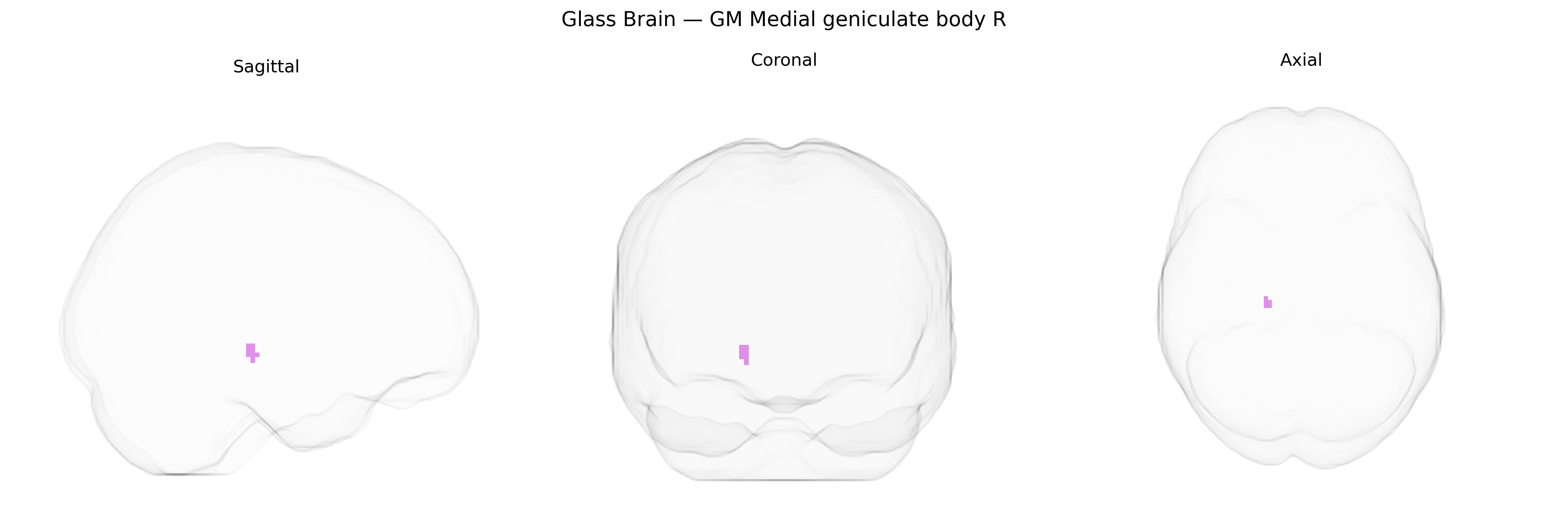
The bilateral GM Medial geniculate body R, as defined in the Juelich maxprob thr25 2mm Atlas, corresponds to gray matter of the right medial geniculate nucleus of the thalamus, a key auditory relay structure situated in the posterior thalamus adjacent to the inferior colliculus. It receives ascending auditory input primarily from the inferior colliculus and projects to the primary and secondary auditory cortices in the temporal lobe, thereby participating in the processing and integration of acoustic information, including sound localization, frequency discrimination, and aspects of auditory attention. The medial geniculate body comprises multiple subnuclei (e.g., ventral, dorsal, and magnocellular divisions) that differ in cytoarchitecture, connectivity, and functional specialization, and serves as an essential node in the central auditory pathway, modulating auditory signals before they reach cortical areas. Medial geniculate nucleus
The medial geniculate body (MGB), the thalamic auditory relay nucleus corresponding to the “GM Medial geniculate body R” region in the Juelich maxprob thr25 2mm atlas, has limited direct, region-specific genetic association data, but several GWAS and imaging–genetics studies implicate thalamic auditory pathways—including the MGB—in broader neuropsychiatric and sensory phenotypes. Variants in genes involved in glutamatergic and GABAergic signaling (such as GRIN2A, GAD1, and others) and in synaptic plasticity and axonal guidance pathways have been associated with thalamic volume and connectivity, which encompass the auditory thalamus and may indirectly affect MGB structure and function. Thalamic nuclei, including auditory components, have been implicated in GWAS of schizophrenia, bipolar disorder, and major depression through polygenic risk scores that correlate with thalamic morphometry and functional connectivity, though these findings usually do not resolve to the MGB specifically. Genetic influences on tinnitus and hyperacusis have been reported in family and twin studies and candidate-gene work (e.g., genes related to synaptic transmission and ion channels), and neuroimaging of tinnitus frequently shows altered MGB activity, suggesting a genetically modulated vulnerability of this region in auditory phantom perception. Additionally, rare variants and copy-number changes linked to developmental auditory pathway disorders and some forms of epilepsy can affect thalamocortical circuitry that includes the MGB. Overall, current genetic evidence points to polygenic, circuit-level influences on thalamic auditory nuclei rather than robust, MGB-specific GWAS hits, with associations mainly inferred through broader thalamic or auditory system imaging–genetics findings and disorder-related circuitry studies.
Overview generated by GPT-4o (2026).
Region ID: 106
Hemisphere: bilateral
Atlas: Juelich maxprob thr25 2mm
Full Quality Version: Download MP4
Full Quality Version: Download MP4
Wali Sidiqyar*, Gaurav Rudravaram*, Elyssa M. McMaster, Trent M. Schwartz, Adam M. Saunders, Kurt G. Schilling, Bennett A. Landman "Introducing SPINS: A Shared Public Visualization Library of Neuroanatomical Structures." Medical Imaging with Deep Learning- short paper
This resource is licensed under CC0 1.0 Universal (Public Domain).