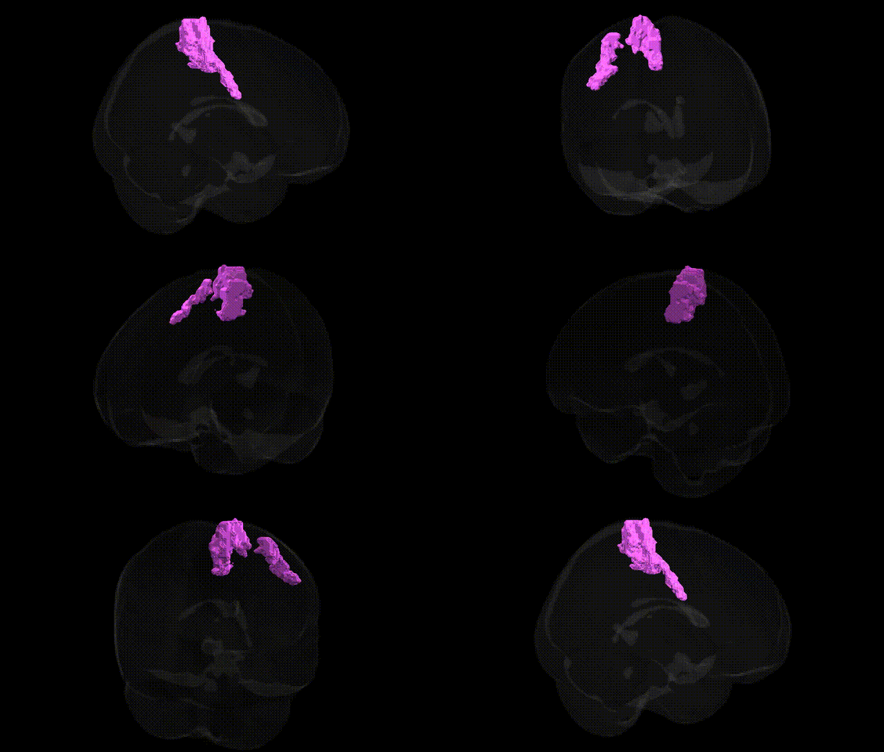
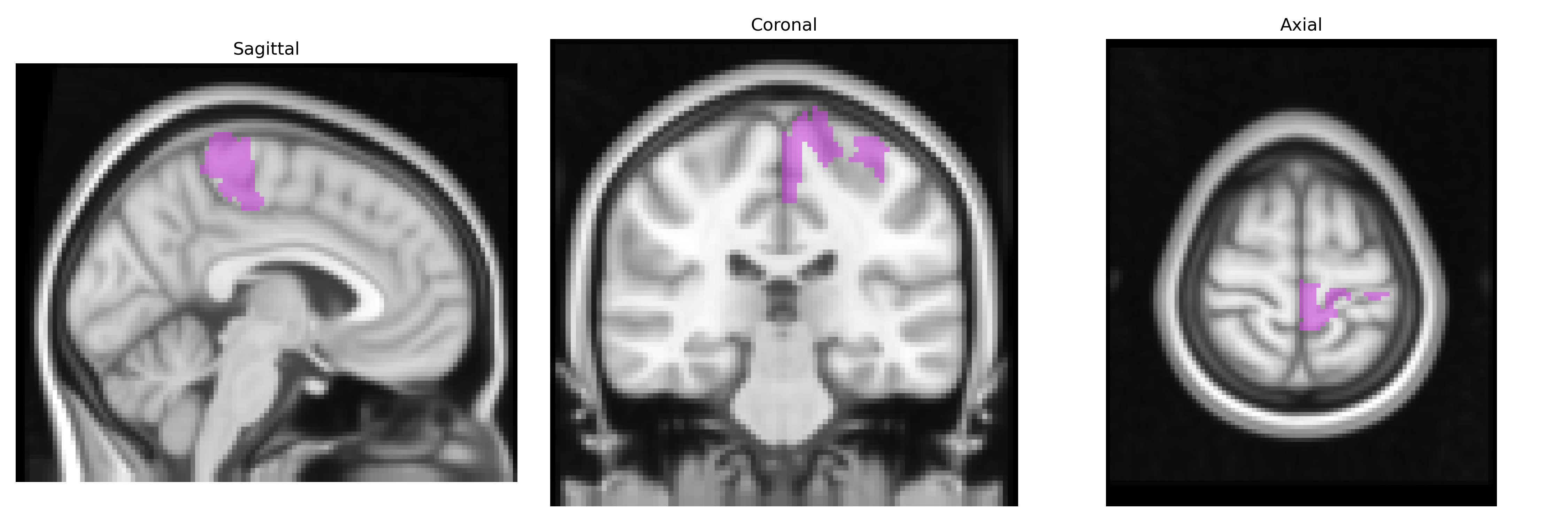
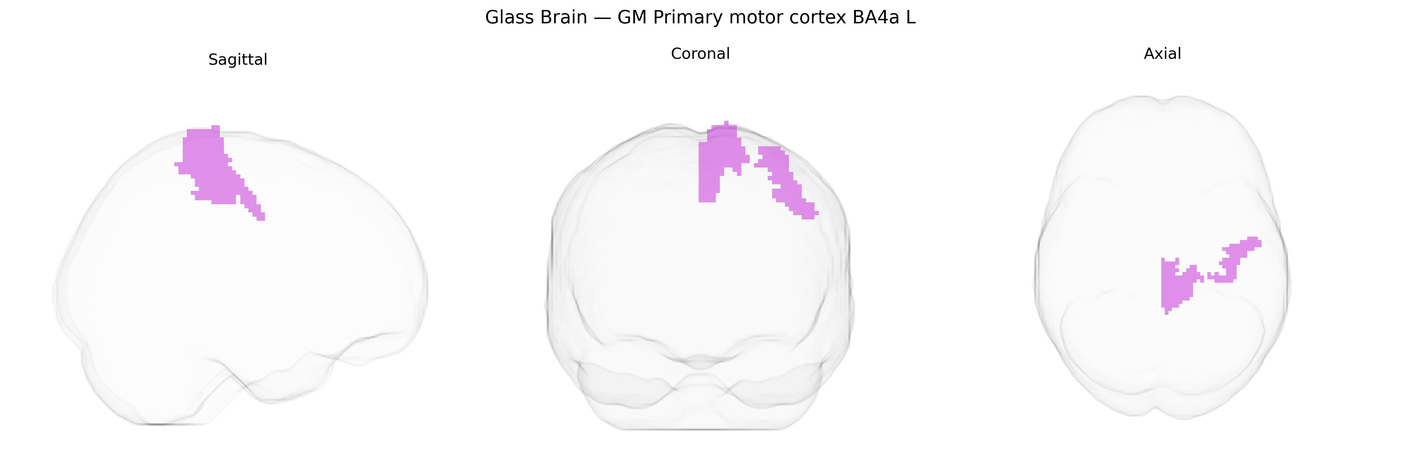
The bilateral GM Primary motor cortex BA4a L from the Juelich maxprob thr25 2mm Atlas corresponds to the agranular portion of Brodmann area 4 in the left hemisphere, located in the precentral gyrus of the frontal lobe and forming a core part of the primary motor cortex. This region contains large pyramidal neurons (including Betz cells) that give rise to corticospinal and corticobulbar projections, mediating voluntary execution of fine motor movements, particularly of the contralateral side of the body. BA4a is distinguished cytoarchitectonically and functionally from adjacent premotor and supplementary motor areas, showing strong somatotopic organization and tight integration with sensorimotor networks for movement planning, force control, and proprioceptive feedback. There is no direct Wikipedia article for “BA4a,” but it is encompassed within the primary motor cortex: Primary motor cortex.
The bilateral primary motor cortex (BA4a, left hemisphere) in the Juelich maxprob thr25 2 mm atlas has been implicated indirectly in multiple genetic and GWAS-based findings, although relatively few studies target this specific cytoarchitectonic subfield. Polygenic risk for neurodevelopmental and neuropsychiatric disorders such as autism spectrum disorder, schizophrenia, and attention-deficit/hyperactivity disorder has been associated with altered cortical thickness, surface area, or activation of primary motor regions, including portions of BA4; implicated genes and loci often involve synaptic function (e.g., GRIN2B, CACNA1C), neurodevelopmental pathways (e.g., CNTNAP2, NRG1), and cytoskeletal or axonal guidance genes. GWAS of motor-related traits—such as reaction time, grip strength, and general motor coordination—have linked variants in genes related to corticospinal tract development and myelination (e.g., loci near BDNF, ROBO/SLIT pathways, and oligodendrocyte-related genes) to structural and functional measures in primary motor cortex. Imaging–genetics studies using large cohorts (e.g., UK Biobank) have identified associations between common variants influencing global brain morphology and regional metrics in BA4a, with motor cortex thickness and surface area showing modest heritability and sharing polygenic architecture with height, educational attainment, and general cognitive ability. Additionally, monogenic movement disorders (such as hereditary spastic paraplegias and some dystonias) frequently show degeneration or functional abnormalities in corticospinal neurons whose cell bodies reside in BA4a, with implicated genes including SPAST, ATL1, and KIF5A; however, these associations are typically defined at the systems level (corticospinal tract and motor cortex broadly) rather than being uniquely localized to the Juelich-defined BA4a L region.
Overview generated by GPT-4o (2026).
Region ID: 47
Hemisphere: bilateral
Atlas: Juelich maxprob thr25 2mm
Full Quality Version: Download MP4
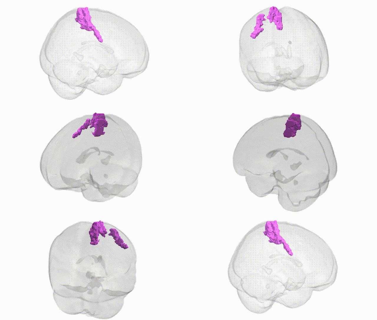
Full Quality Version: Download MP4
Wali Sidiqyar*, Gaurav Rudravaram*, Elyssa M. McMaster, Trent M. Schwartz, Adam M. Saunders, Kurt G. Schilling, Bennett A. Landman "Introducing SPINS: A Shared Public Visualization Library of Neuroanatomical Structures." Medical Imaging with Deep Learning- short paper
This resource is licensed under CC0 1.0 Universal (Public Domain).