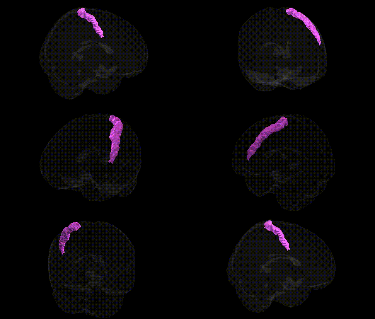
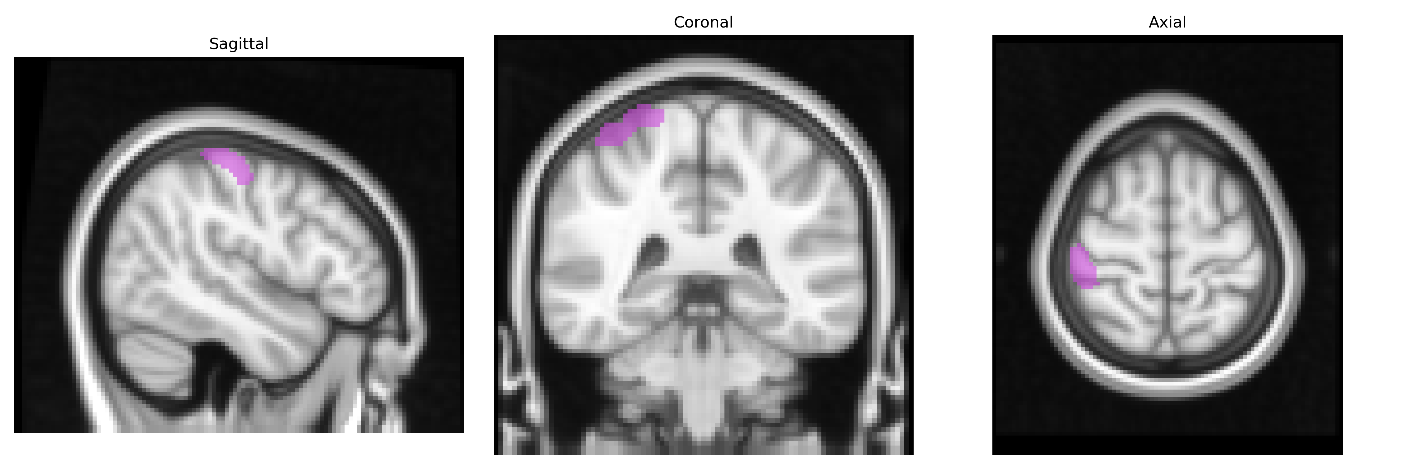
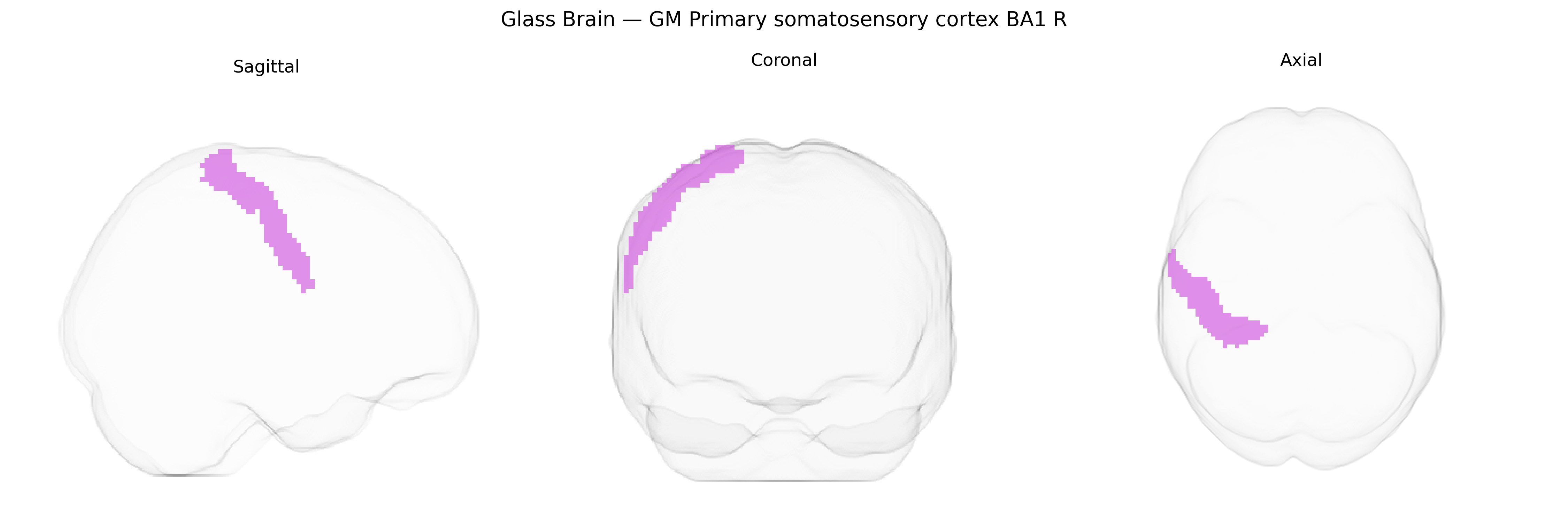
The bilateral GM Primary somatosensory cortex BA1 R from the Juelich maxprob thr25 2mm Atlas corresponds to Brodmann area 1 within the postcentral gyrus of the parietal lobe, primarily located on the crown of the gyrus posterior to the central sulcus. This region forms part of the primary somatosensory cortex and is heavily involved in processing tactile information, especially texture and fine-grained features of objects, integrating input from rapidly adapting cutaneous mechanoreceptors. BA1 receives afferent projections from thalamocortical pathways (notably from the ventral posterior nucleus of the thalamus) and from adjacent somatosensory areas (BA3 and BA2), and contributes to ascending somatosensory processing streams that support conscious perception of touch and tactile discrimination. Although there is no dedicated Wikipedia page specifically for Brodmann area 1 of the primary somatosensory cortex, it is encompassed within the broader description of the Primary somatosensory cortex.
The bilateral gray matter primary somatosensory cortex BA1 (right hemisphere) from the Juelich maxprob thr25 2mm atlas has been implicated in genetic studies largely through variants influencing cortical structure and pain-related processing rather than region-specific GWAS hits. Neuroimaging genetics consortia such as ENIGMA and UK Biobank have identified common variants in genes involved in neurodevelopment and synaptic function (e.g., microtubule and axon guidance genes like TUBB, growth signaling pathways such as those involving BDNF and IGF1, and broad neurodevelopmental loci including genes near MAPT and DCC) that are associated with global or parietal cortical thickness and surface area, which encompass BA1 regions. GWAS of chronic pain, somatosensory phenotypes, and neuropathic pain have linked polygenic risk scores and specific loci (for instance, variants in SCN9A and other nociception-related genes) to functional and structural alterations in somatosensory cortices, suggesting a genetically mediated modulation of sensory-discriminative pain processing in BA1. Additionally, genetic risk for neurodevelopmental disorders (such as schizophrenia and autism spectrum conditions) and certain cognitive traits has been associated with altered parietal and somatosensory cortex morphology and connectivity in imaging–genetics studies, indirectly implicating BA1 through spatially overlapping structural and functional changes, although no high-confidence GWAS signals are known that specifically and uniquely map to the right BA1 field in the Juelich atlas.
Overview generated by GPT-4o (2026).
Region ID: 52
Hemisphere: bilateral
Atlas: Juelich maxprob thr25 2mm
Full Quality Version: Download MP4
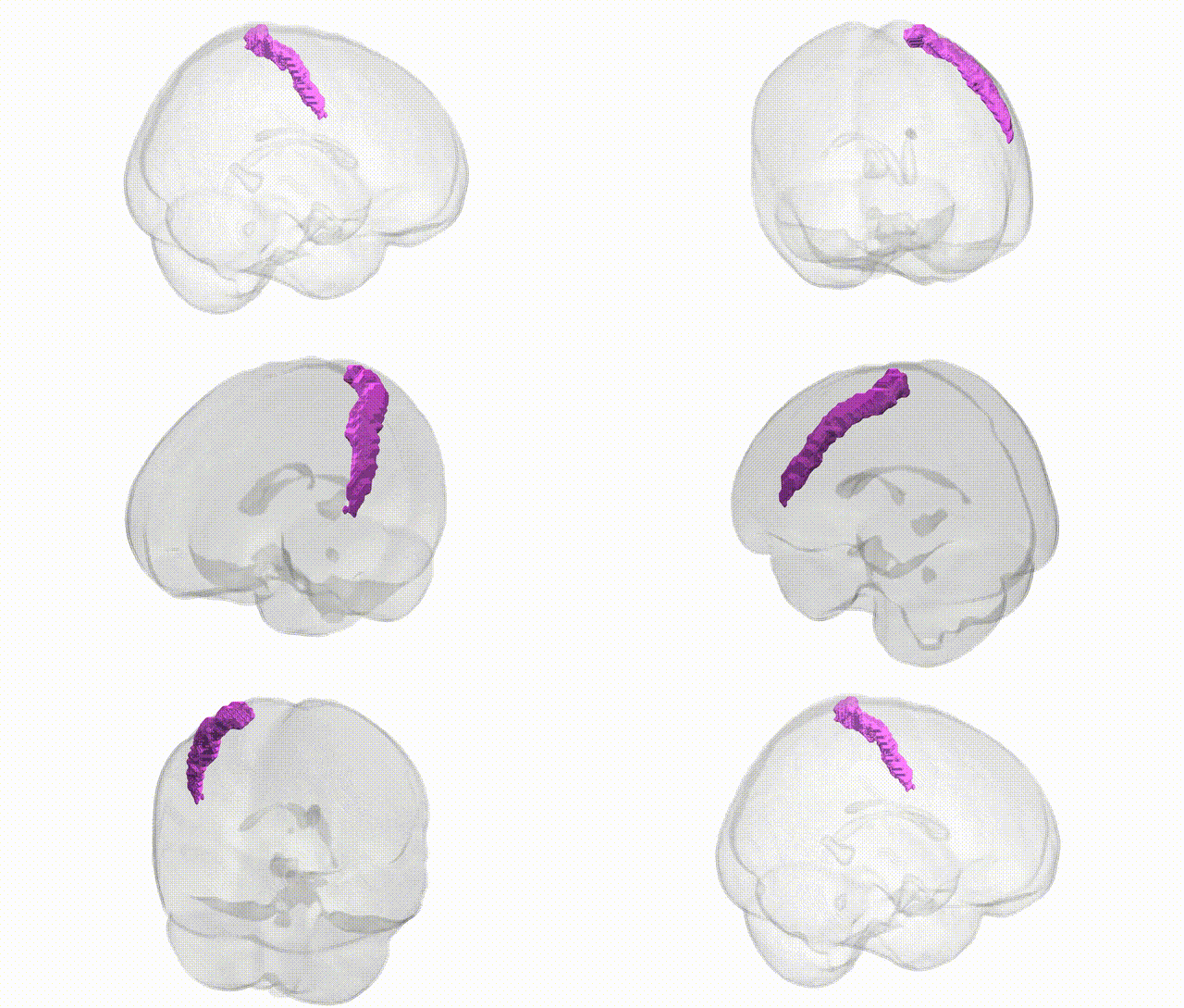
Full Quality Version: Download MP4
Wali Sidiqyar*, Gaurav Rudravaram*, Elyssa M. McMaster, Trent M. Schwartz, Adam M. Saunders, Kurt G. Schilling, Bennett A. Landman "Introducing SPINS: A Shared Public Visualization Library of Neuroanatomical Structures." Medical Imaging with Deep Learning- short paper
This resource is licensed under CC0 1.0 Universal (Public Domain).