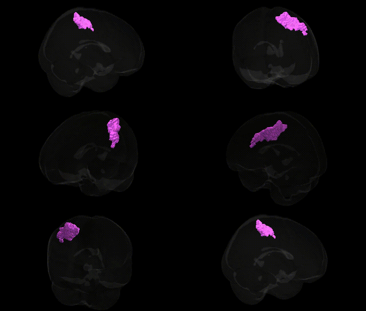
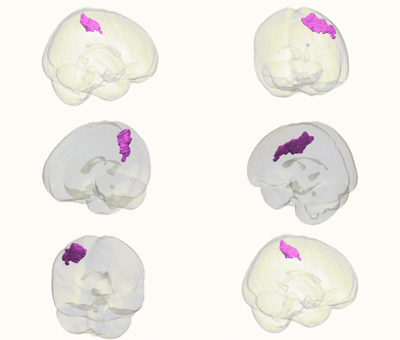
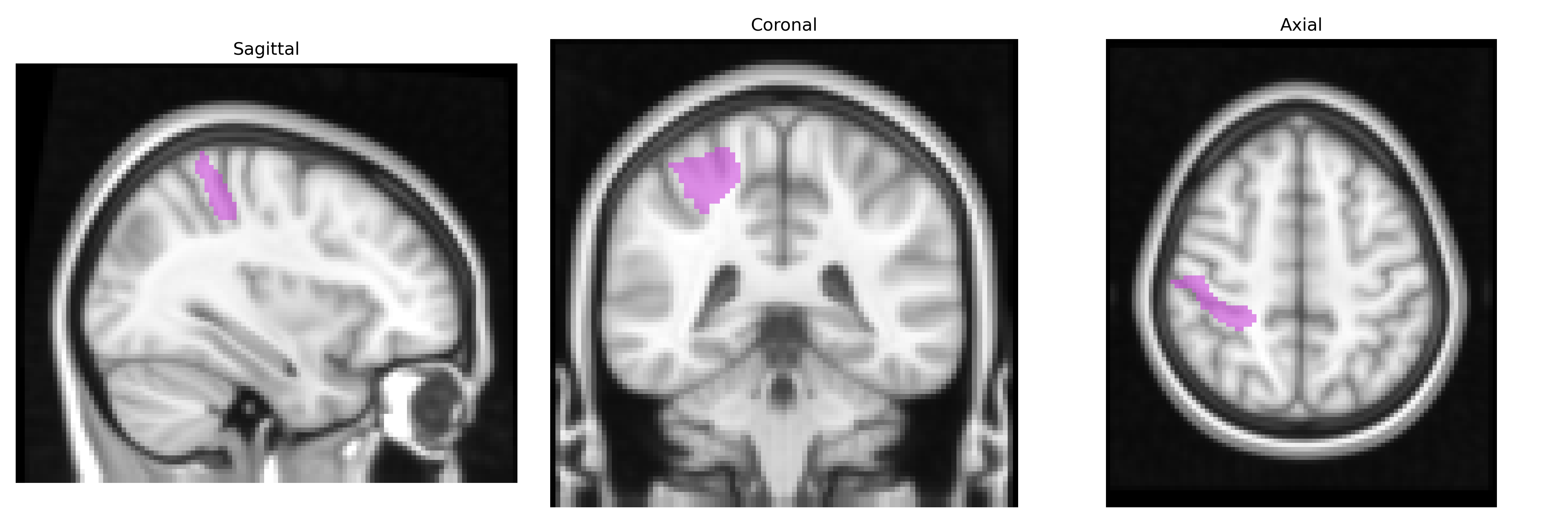
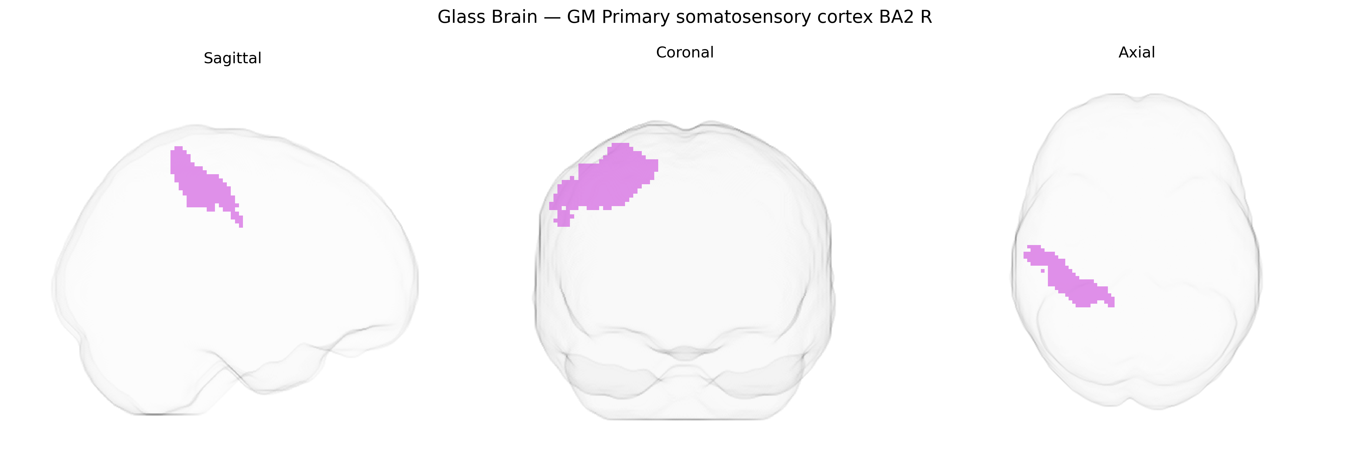
The bilateral GM Primary somatosensory cortex BA2 R, as defined in the Juelich maxprob thr25 2mm atlas, corresponds to Brodmann area 2 within the postcentral gyrus of the parietal lobe, predominantly in the right hemisphere but represented bilaterally as a gray-matter functional region. BA2 is part of the primary somatosensory cortex and is involved in higher-order processing of tactile and proprioceptive information, integrating signals from cutaneous and deep receptors to support complex features such as size, shape, texture, and limb position perception. It receives input from other somatosensory areas (including BA3 and BA1) and contributes to sensorimotor integration necessary for fine object manipulation and coordinated movement. There is no direct Wikipedia article specifically for “Brodmann area 2”; a closely related and encompassing structure is the primary somatosensory cortex: Primary somatosensory cortex.
The bilateral gray matter primary somatosensory cortex BA2 (right hemisphere) from the Juelich maxprob thr25 2mm atlas has been implicated in genetic studies primarily through imaging genetics and GWAS that link cortical structure and sensorimotor processing to common variants. Large-scale GWAS of cortical thickness and surface area (e.g., ENIGMA, UK Biobank) have identified SNPs in genes involved in neurodevelopment, synaptic function, and axon guidance (such as variants near CENPW, TBR1, and genes in Wnt and FGF signaling pathways) that influence somatosensory cortical morphology, though these findings are often reported at lobar or broader parcellation levels rather than specifically BA2 R. Polygenic scores associated with cognitive performance, educational attainment, and general brain volume show correlations with structural measures in parietal and somatosensory regions, suggesting pleiotropic genetic effects on BA2-related circuits. Genetic studies of chronic pain, neuropathic pain, and somatosensory dysfunction (including pain sensitivity GWAS and rare-variant analyses in sodium channel genes such as SCN9A) indirectly implicate BA2 R by demonstrating altered activation or connectivity of this region in carriers of specific alleles, although the associations are typically mediated via peripheral and thalamocortical pathways rather than being region-specific. Neurodevelopmental and psychiatric GWAS, including those for autism spectrum disorder, ADHD, and schizophrenia, have identified risk loci in genes regulating synaptic plasticity and cortical development (e.g., CACNA1C, GRIN2A, CNTNAP2), and imaging-genetic work has shown that risk allele carriers often exhibit atypical somatosensory or parietal cortical organization, linking these genetic variants to functional differences in BA2-related tactile and proprioceptive processing without unique, isolated GWAS hits dedicated exclusively to BA2 R.
Overview generated by GPT-4o (2026).
Region ID: 54
Hemisphere: bilateral
Atlas: Juelich maxprob thr25 2mm
Full Quality Version: Download MP4
Full Quality Version: Download MP4
Wali Sidiqyar*, Gaurav Rudravaram*, Elyssa M. McMaster, Trent M. Schwartz, Adam M. Saunders, Kurt G. Schilling, Bennett A. Landman "Introducing SPINS: A Shared Public Visualization Library of Neuroanatomical Structures." Medical Imaging with Deep Learning- short paper
This resource is licensed under CC0 1.0 Universal (Public Domain).