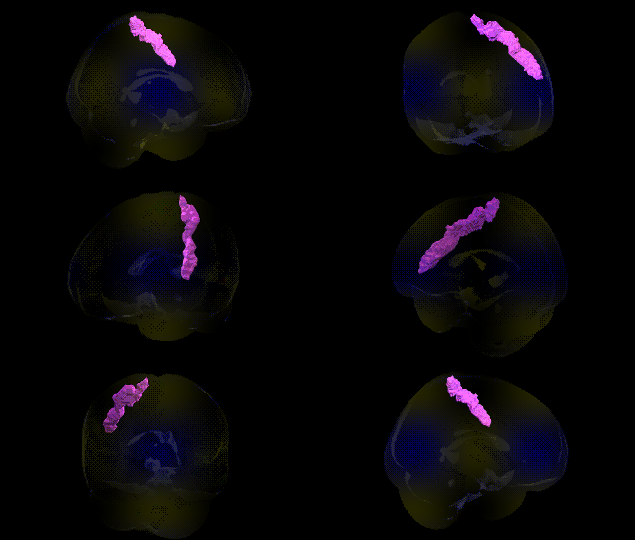
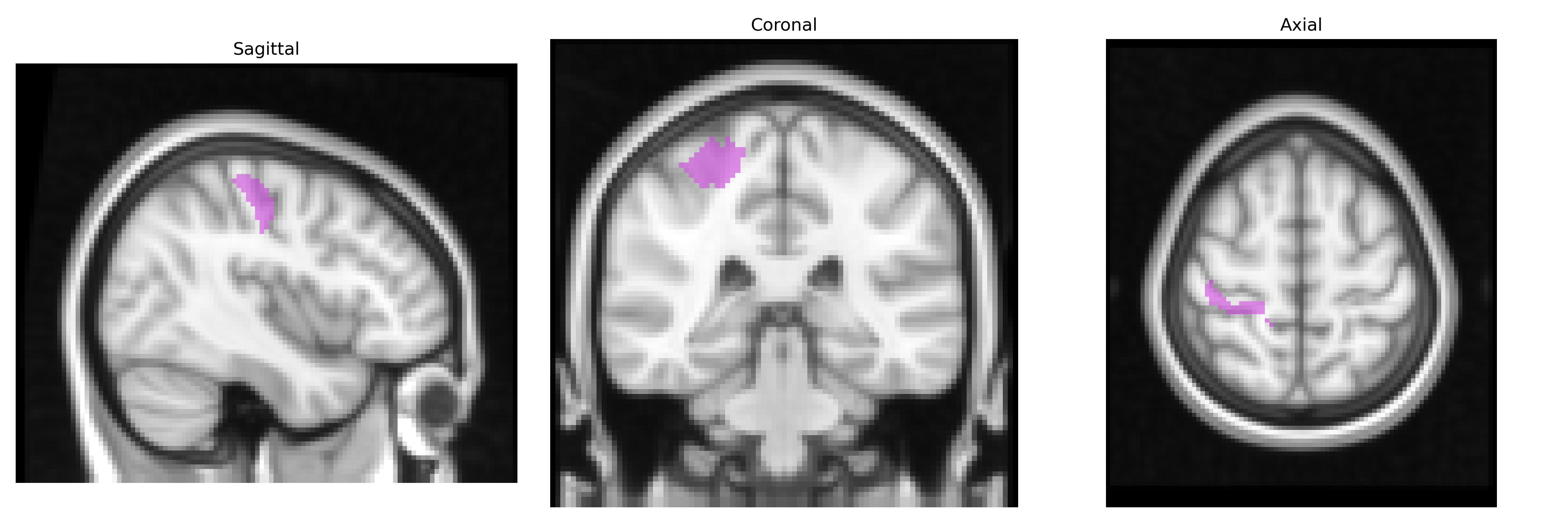
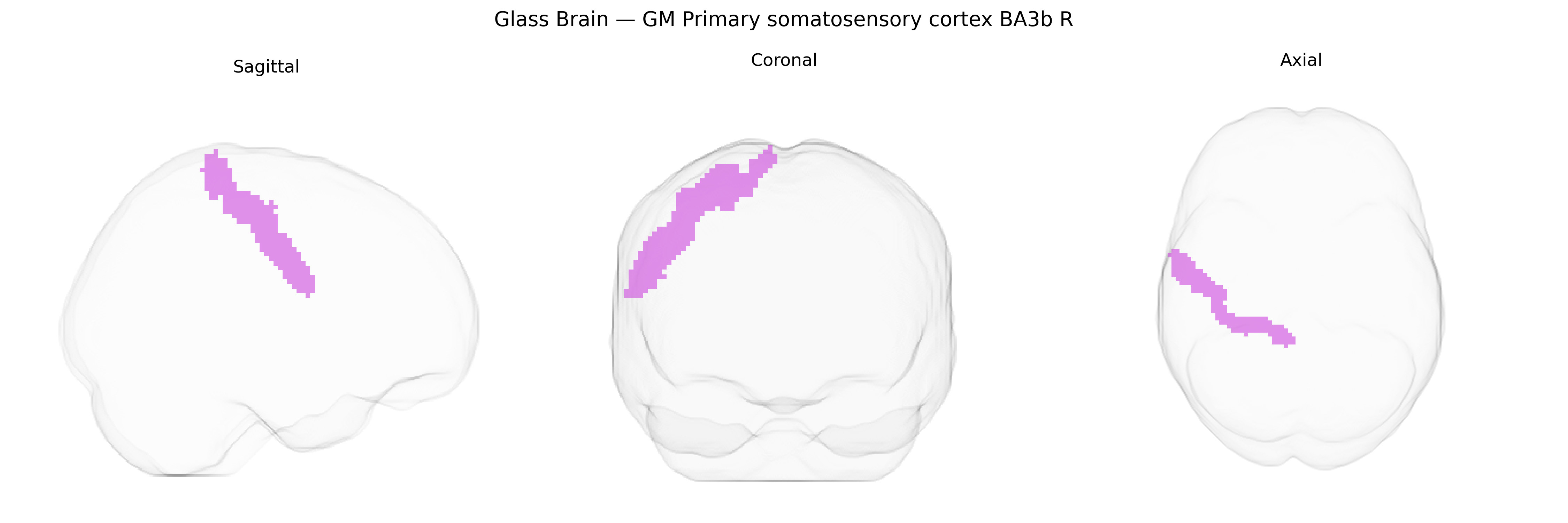
The bilateral GM Primary somatosensory cortex BA3b R, as defined in the Juelich maxprob thr25 2mm atlas, corresponds to the cytoarchitectonically specified Brodmann area 3b located in the postcentral gyrus of the parietal lobe, forming a core component of the primary somatosensory cortex. This region receives dense thalamocortical input from somatosensory relay nuclei and is specialized for processing tactile information, including fine touch, texture, and form, with a highly organized somatotopic representation of the body surface. Its granular cortical architecture (well-developed layer IV) supports precise spatial discrimination and integration of cutaneous signals, and it plays a critical role in conscious perception of touch and in guiding sensorimotor behavior through projections to adjacent somatosensory areas and motor-related regions. There is no dedicated Wikipedia article specifically for “BA3b”; see the related entry for the encompassing structure: Primary somatosensory cortex.
The bilateral GM Primary somatosensory cortex BA3b R region in the Juelich maxprob thr25 2mm Atlas corresponds to a core area of the postcentral gyrus involved in basic tactile and proprioceptive processing, and genetic associations involving this territory are typically inferred from broader imaging-genetics and GWAS studies of cortical thickness, surface area, and somatotopic sensorimotor regions rather than BA3b-specific analyses. Large-scale imaging GWAS consortia (e.g., ENIGMA, UK Biobank) have identified common variants in genes related to neurodevelopment, synaptic function, and axon guidance (such as variants near KIAA0586, MIR137, MEF2C, and others) associated with structural metrics of the postcentral gyrus or global somatosensory cortex, suggesting polygenic influences on this region’s morphology and connectivity. Somatosensory cortex–adjacent loci have been implicated in neurodevelopmental and neuropsychiatric disorders—particularly autism spectrum disorder, schizophrenia, ADHD, and epilepsy—where risk variants affect cortical organization and sensory processing, though these links generally span multiple nearby regions and are not exclusive to BA3b. GWAS of chronic pain, neuropathic pain, and somatosensory phenotypes (e.g., pain sensitivity, tactile thresholds) have also highlighted variants in genes involved in ion channels, inflammatory signaling, and neuroplasticity (such as SCN9A and other channel genes), with imaging-genetics work showing that these variants can modulate somatosensory cortical activation or structure. However, current evidence for direct, region-specific genetic associations with the Juelich-defined BA3b R field remains limited, and most genetic findings relate to broader postcentral or sensorimotor territories rather than precisely to bilateral GM Primary somatosensory cortex BA3b R as delineated by this atlas.
Overview generated by GPT-4o (2026).
Region ID: 58
Hemisphere: bilateral
Atlas: Juelich maxprob thr25 2mm
Full Quality Version: Download MP4
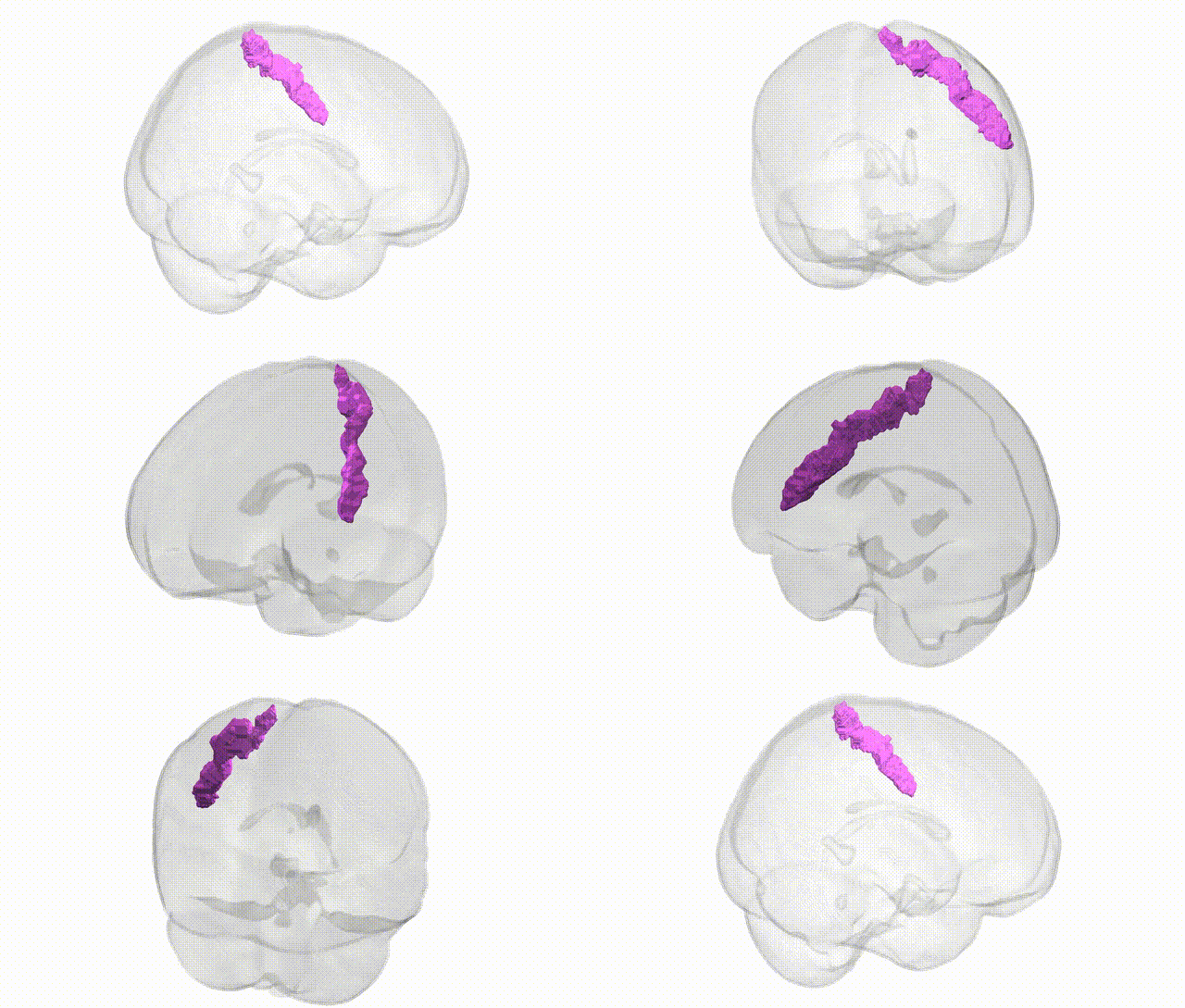
Full Quality Version: Download MP4
Wali Sidiqyar*, Gaurav Rudravaram*, Elyssa M. McMaster, Trent M. Schwartz, Adam M. Saunders, Kurt G. Schilling, Bennett A. Landman "Introducing SPINS: A Shared Public Visualization Library of Neuroanatomical Structures." Medical Imaging with Deep Learning- short paper
This resource is licensed under CC0 1.0 Universal (Public Domain).