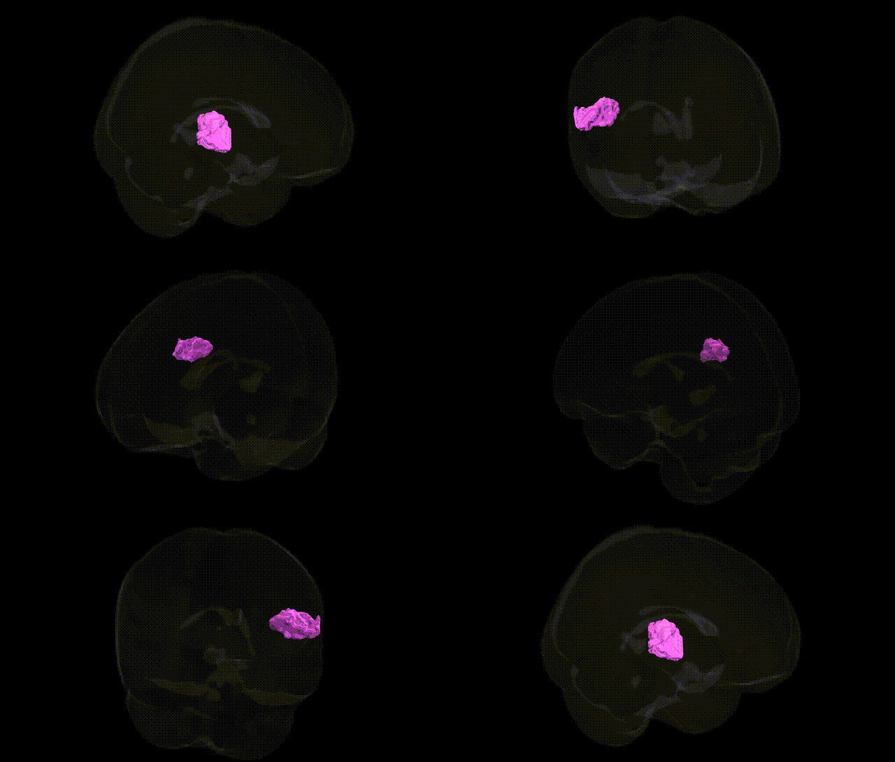
The bilateral GM Secondary somatosensory cortex Parietal operculum OP1 L, as defined in the Juelich maxprob thr25 2mm atlas, corresponds to a cytoarchitectonic subregion of the parietal operculum that forms part of the human secondary somatosensory cortex (SII). This region is located on the upper bank of the lateral sulcus within the inferior parietal area, receiving dense somatosensory input from primary somatosensory cortex and thalamic nuclei, and is implicated in higher-order tactile processing, including texture discrimination, integration of bilateral somatosensory information, and aspects of pain and sensorimotor integration. OP1 exhibits multimodal convergence, interacting with insular, frontal, and parietal association areas, and contributes to the perceptual and affective dimensions of touch and nociception. There is no direct Wikipedia article for OP1; a closely related structure is the Secondary somatosensory cortex.
The bilateral GM Secondary somatosensory cortex Parietal operculum OP1 (L) from the Juelich maxprob thr25 2 mm atlas lies in the posterior parietal operculum and overlaps core human SII, a region repeatedly implicated in genetically influenced somatosensory, pain, and sensorimotor traits. Twin and heritability studies show substantial genetic contributions to cortical thickness and surface area in parietal opercular and SII territories, and GWAS of brain morphology have linked common variants near genes involved in neurodevelopment (for example, microtubule- and axon-guidance–related genes) to inter-individual differences in parietal operculum structure, although specific OP1-only associations are rarely isolated. Functional imaging genetics and polygenic score work indicate that alleles conferring risk for chronic pain conditions (such as migraine, neuropathic and musculoskeletal pain) and related traits (pain sensitivity, nociception, and affective pain processing) modulate activation and connectivity in SII/OP1, consistent with its role in encoding stimulus intensity and localization. Genetic risk for autism spectrum disorder, schizophrenia, and attention-deficit/hyperactivity disorder has been associated in large imaging-genetics cohorts with altered somatosensory and parietal opercular anatomy and functional response profiles, suggesting pleiotropic effects of neurodevelopmental risk loci on SII circuitry; however, these associations typically involve broader peri-sylvian sensorimotor networks, so the contribution of OP1 itself is inferred rather than anatomically specific.
Overview generated by GPT-4o (2026).
Region ID: 59
Hemisphere: bilateral
Atlas: Juelich maxprob thr25 2mm
Full Quality Version: Download MP4
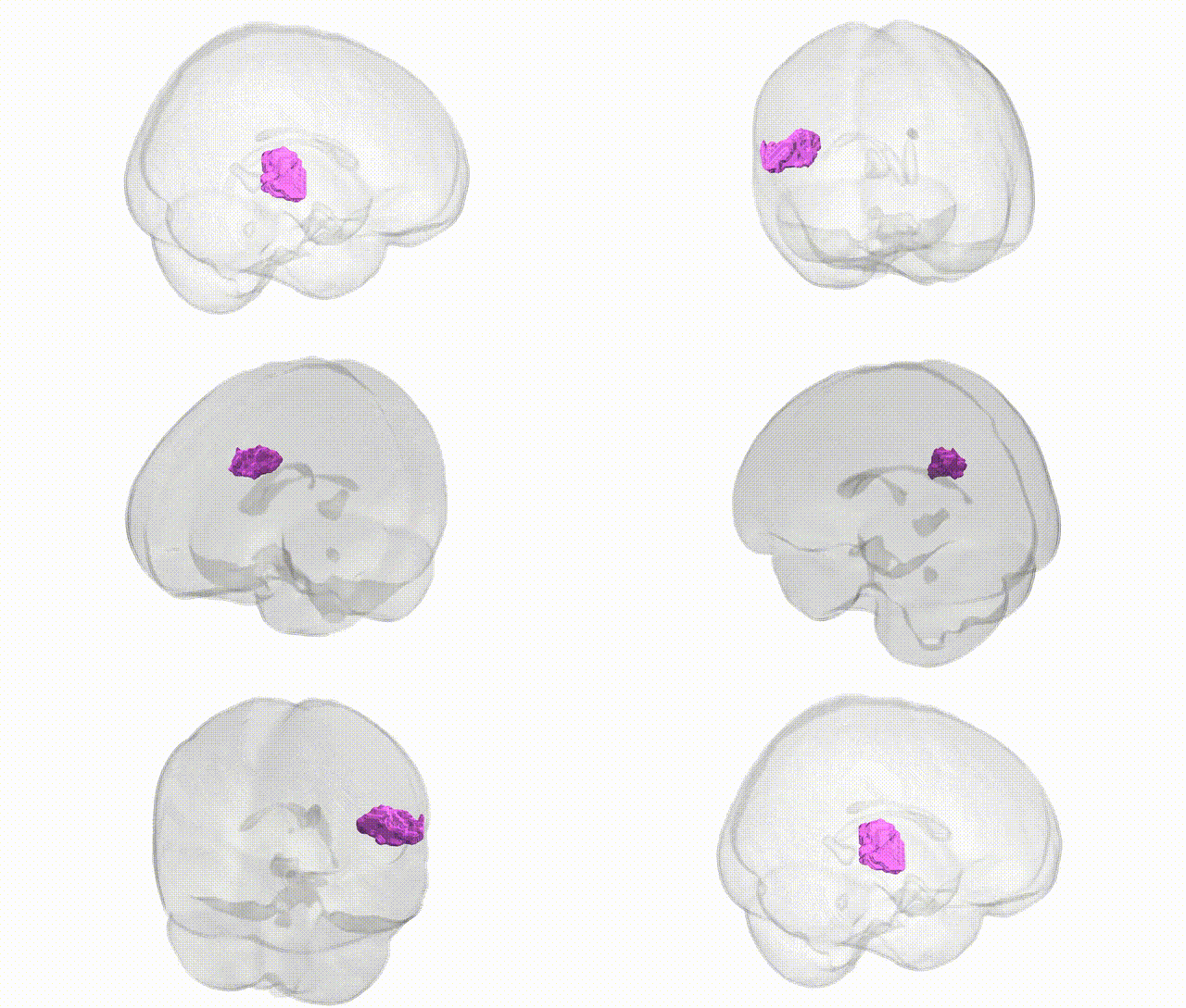
Full Quality Version: Download MP4
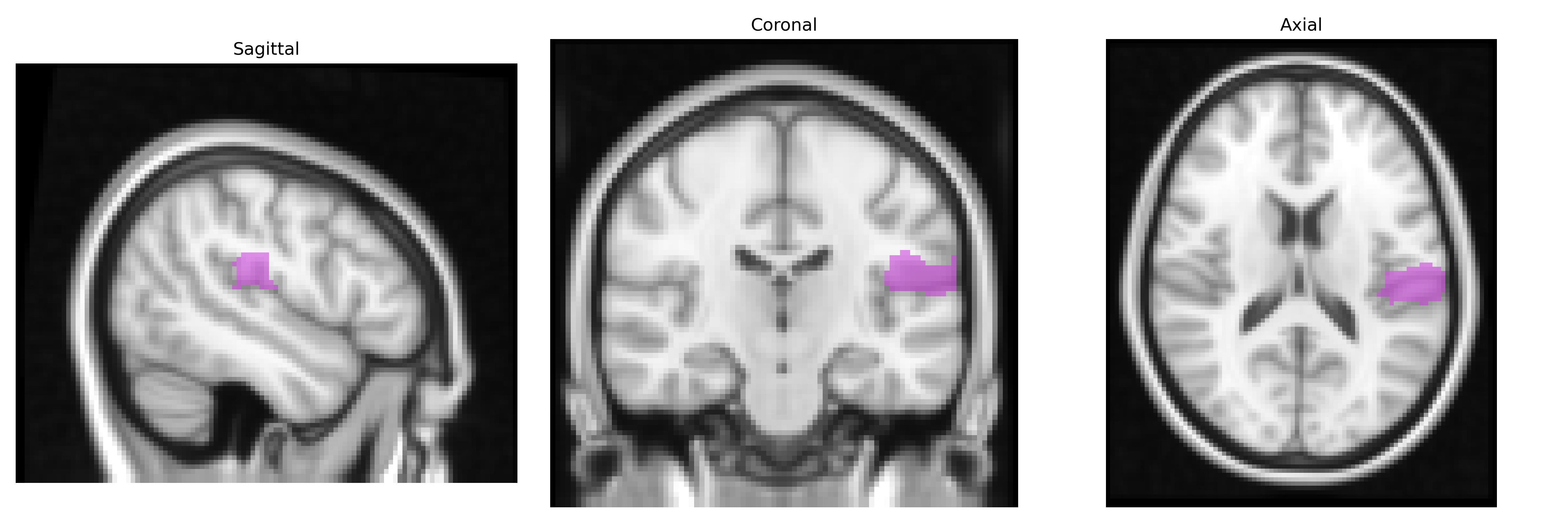
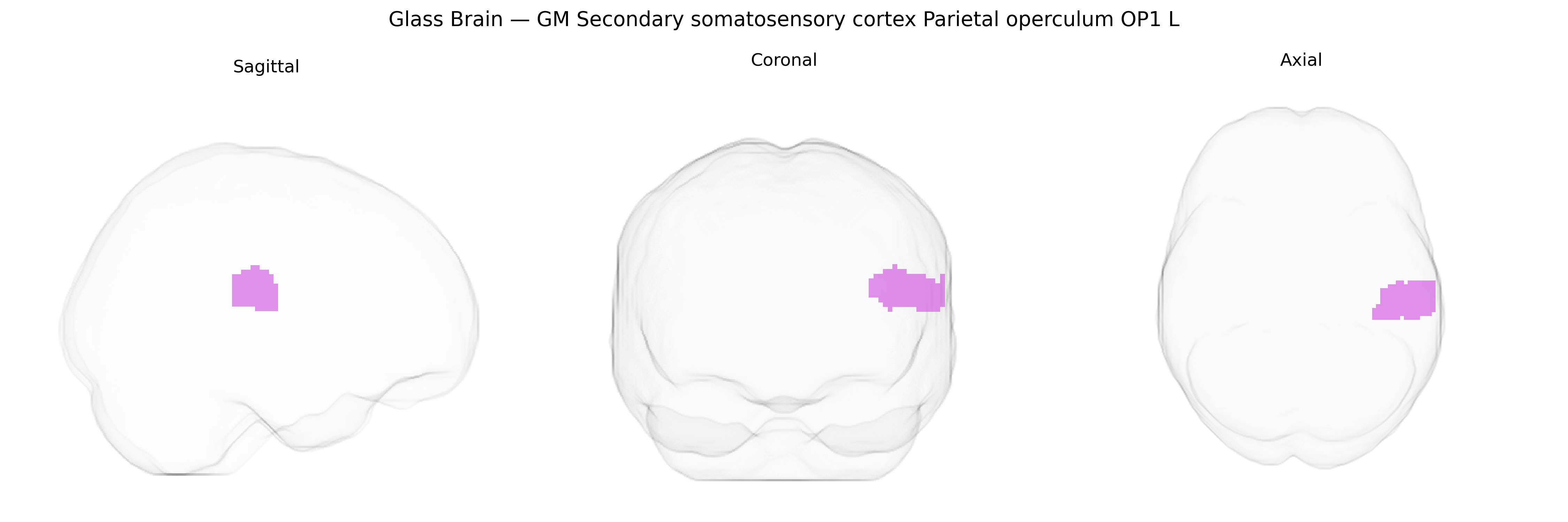
Wali Sidiqyar*, Gaurav Rudravaram*, Elyssa M. McMaster, Trent M. Schwartz, Adam M. Saunders, Kurt G. Schilling, Bennett A. Landman "Introducing SPINS: A Shared Public Visualization Library of Neuroanatomical Structures." Medical Imaging with Deep Learning- short paper
This resource is licensed under CC0 1.0 Universal (Public Domain).