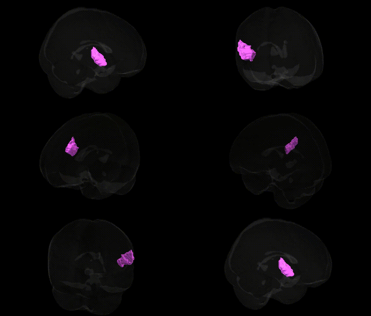
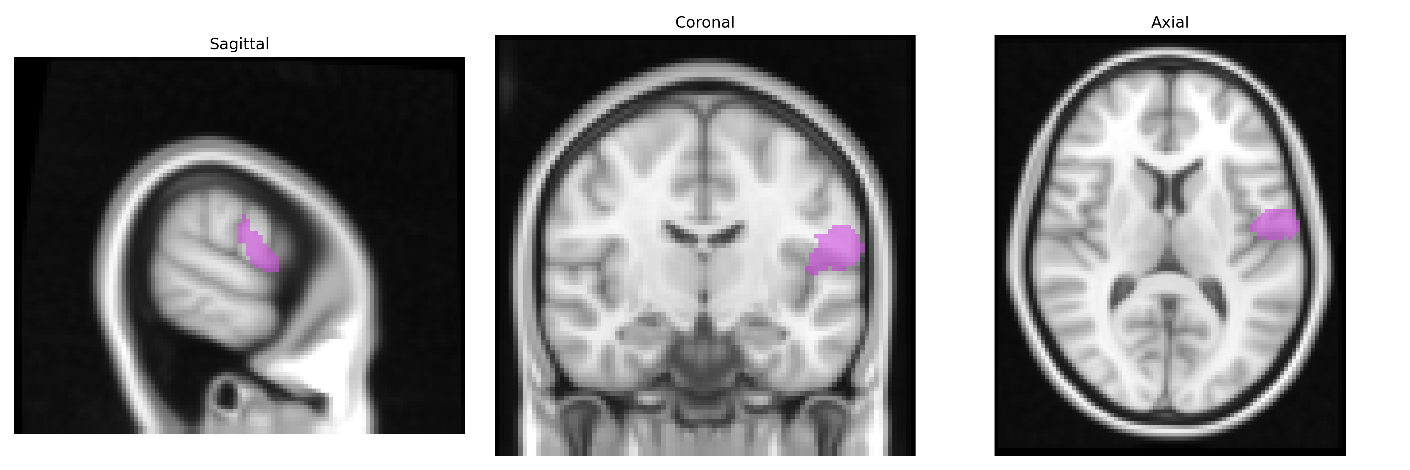
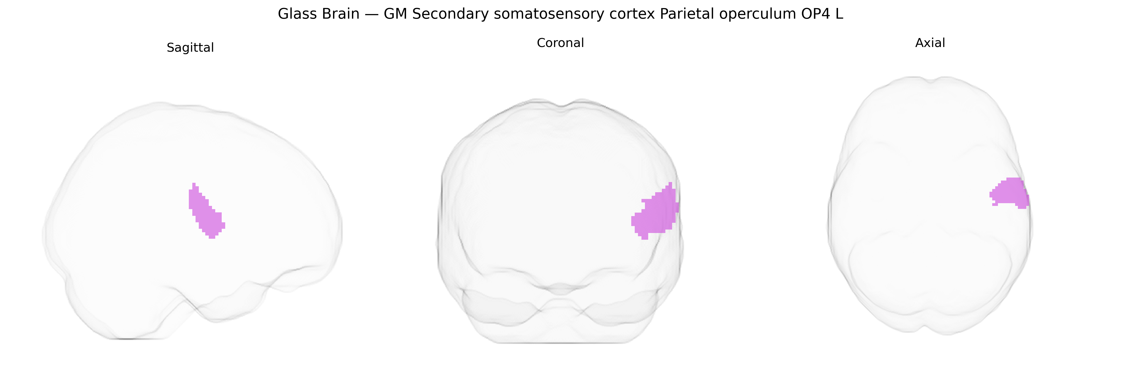
The bilateral GM Secondary somatosensory cortex Parietal operculum OP4 L, as defined in the Juelich maxprob thr25 2mm atlas, corresponds to a cytoarchitectonically distinct subregion of the parietal operculum that forms part of the secondary somatosensory cortex (SII). Located in the lateral sulcus overlying the insula, OP4 is involved in higher-order processing of tactile and proprioceptive information, including integration of bilateral somatosensory inputs, texture discrimination, and aspects of sensorimotor integration relevant for object manipulation and body representation. Neuronal populations in OP4 show multimodal responsiveness, receiving convergent input from primary somatosensory cortex and other association areas, and contribute to the transformation of basic somatosensory signals into more complex perceptual and cognitive representations. There is no direct Wikipedia article for OP4; a closely related and encompassing structure is the Parietal operculum.
The bilateral GM Secondary somatosensory cortex Parietal operculum OP4 (L) region, as defined in the Juelich maxprob thr25 2 mm atlas, falls within the parietal operculum/SII, a multimodal somatosensory area for which genetic associations are typically inferred from broader imaging-genetics and GWAS-based neuroimaging studies rather than OP4-specific analyses; large-scale efforts such as ENIGMA and UK Biobank implicate this region’s microstructure and cortical morphology in heritable variation related to somatosensory processing, pain perception, motor-sensory integration, and tactile discrimination, with polygenic influences overlapping genes involved in neurodevelopment, synaptic organization, and myelination (e.g., general cortical surface area and thickness loci including variants near genes such as MIR588, THBS1, and others reported for parietal and opercular cortices). GWAS and imaging-genetics studies link parietal operculum and adjacent SII regions to chronic pain conditions (including neuropathic and musculoskeletal pain), migraine, and trait pain sensitivity, in which risk variants often converge on neurotransmission (e.g., glutamatergic and GABAergic pathways) and neuroinflammatory signaling, although these are not uniquely assigned to OP4. In neurodevelopmental and psychiatric conditions such as autism spectrum disorder, ADHD, and schizophrenia, genetic risk scores and disorder-associated variants have been associated with altered structure and functional connectivity in secondary somatosensory and parietal opercular territories, reflecting pleiotropic genetic influences on sensorimotor integration and body representation, yet specific OP4-localized genetic signals remain rare due to current atlas and parcellation resolutions. Overall, existing genetic evidence points to polygenic, distributed influences on the broader parietal operculum/SII complex rather than discrete OP4-specific loci, with associations spread across neurodevelopmental, pain, and psychiatric trait architectures.
Overview generated by GPT-4o (2026).
Region ID: 65
Hemisphere: bilateral
Atlas: Juelich maxprob thr25 2mm
Full Quality Version: Download MP4
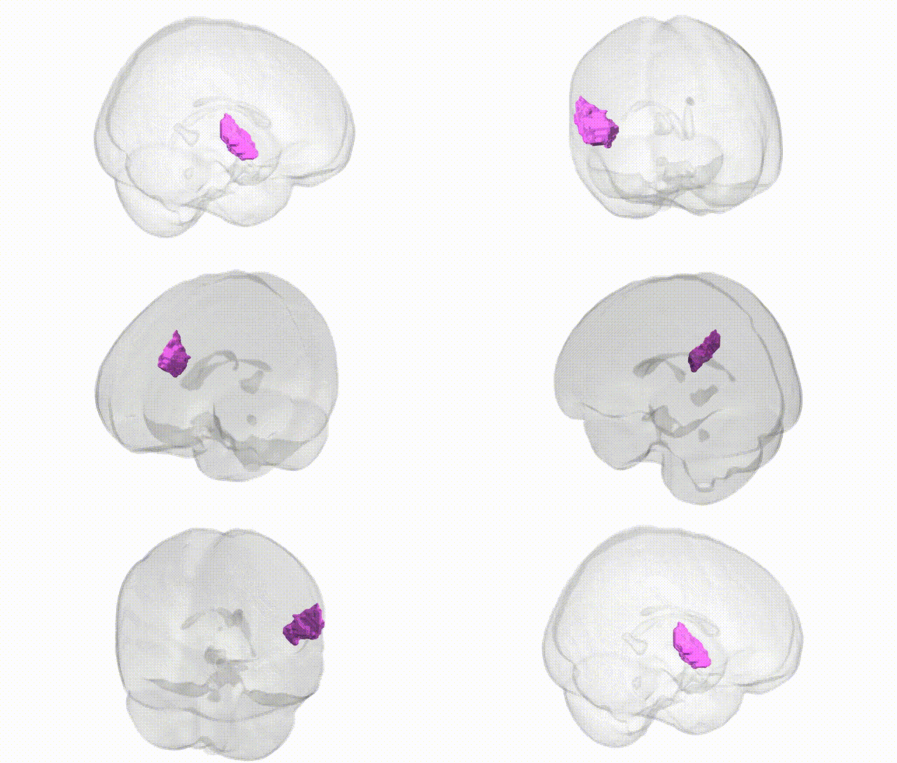
Full Quality Version: Download MP4
Wali Sidiqyar*, Gaurav Rudravaram*, Elyssa M. McMaster, Trent M. Schwartz, Adam M. Saunders, Kurt G. Schilling, Bennett A. Landman "Introducing SPINS: A Shared Public Visualization Library of Neuroanatomical Structures." Medical Imaging with Deep Learning- short paper
This resource is licensed under CC0 1.0 Universal (Public Domain).