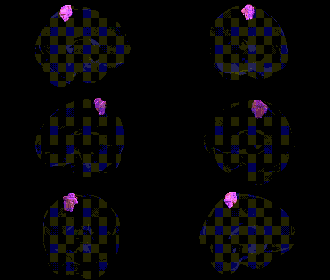
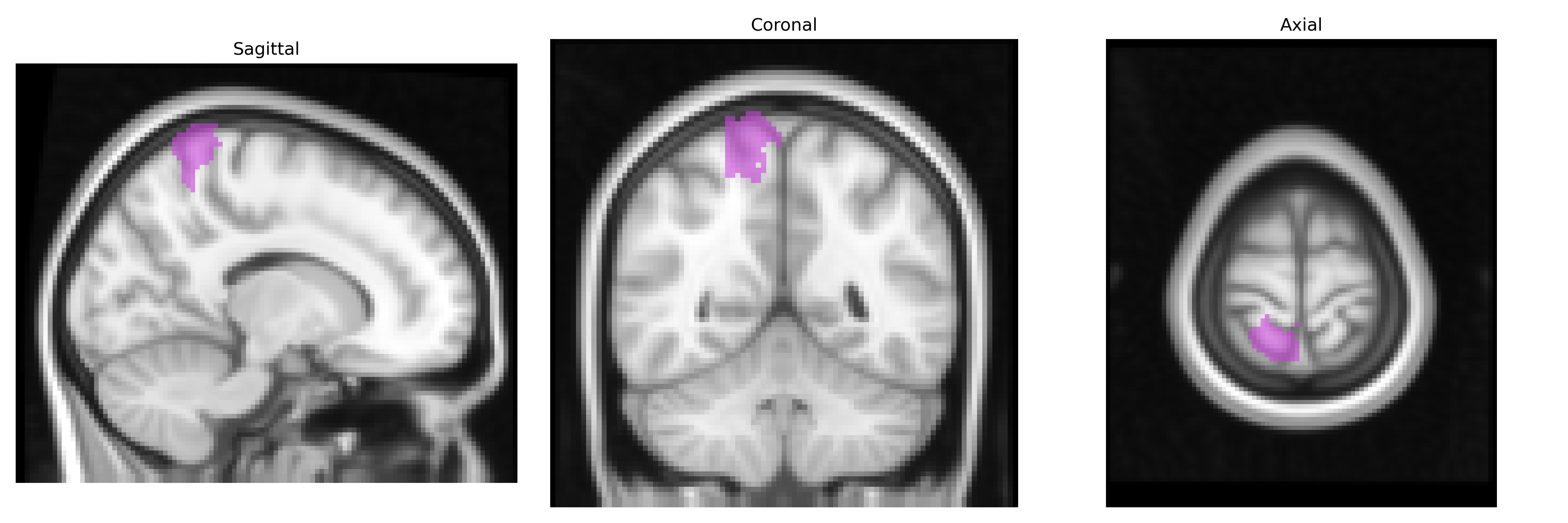
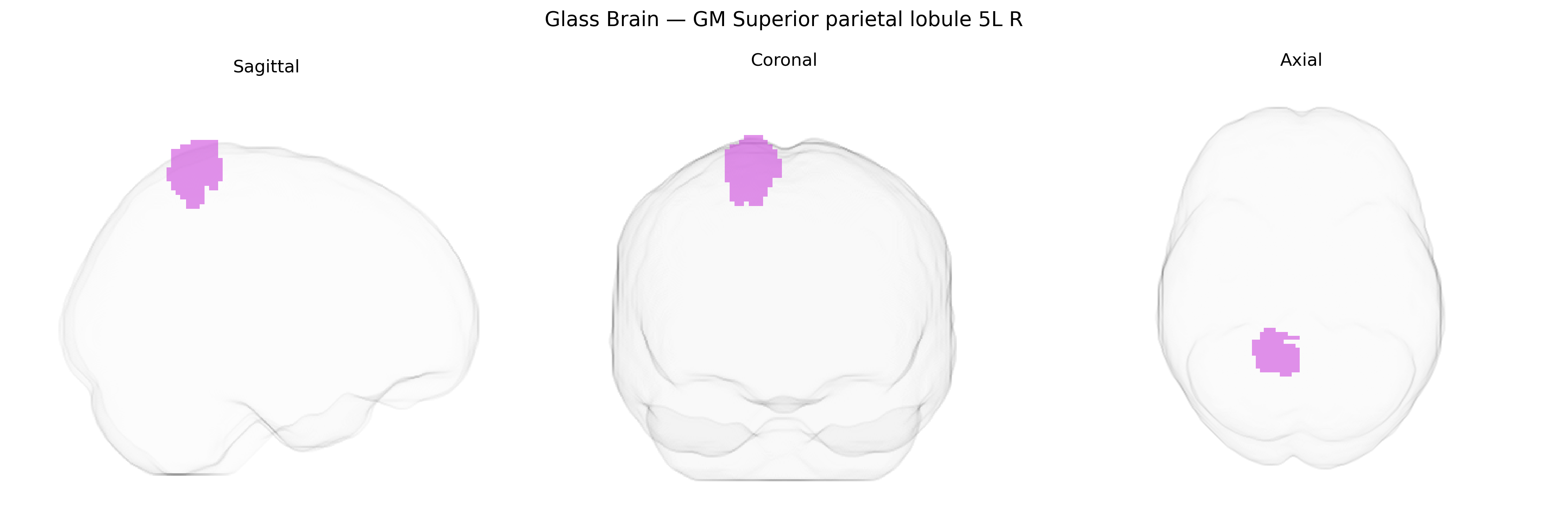
The bilateral GM Superior parietal lobule area 5L (right hemisphere) in the Juelich maxprob thr25 2 mm atlas corresponds to a cytoarchitectonically defined subdivision of Brodmann area 5 within the superior parietal lobule, implicated in higher-order somatosensory and sensorimotor integration. Neurons in this region receive convergent input from primary and secondary somatosensory cortices as well as premotor and visual areas, supporting functions such as multisensory body representation, proprioceptive processing, and the transformation of sensory coordinates into motor commands for reaching and grasping. Functionally, it contributes to spatial attention, coordination of limb movements, and the integration of tactile and proprioceptive information necessary for complex object manipulation and body schema maintenance. There is no direct link specific to “area 5L”; a related structure within which it resides is the Superior parietal lobule.
The bilateral GM Superior parietal lobule (SPL) in the Jülich maxprob thr25 2 mm atlas—approximately corresponding to area 5L/5R in the superior parietal cortex—has been implicated in multiple genetic and GWAS findings, primarily via imaging-genetics studies of cortical thickness, surface area, and functional activation. Large-scale ENIGMA and UK Biobank analyses have identified common variants near genes involved in neurodevelopment, synaptic organization, and axon guidance (for example in pathways including WNT signaling, cell-adhesion molecules, and calcium channel genes) that modulate SPL morphology and connectivity. Polygenic risk for schizophrenia, autism spectrum disorder, and ADHD shows associations with altered superior parietal structure and function, including reduced cortical thickness or atypical activation in tasks involving visuospatial attention, working memory, and sensorimotor integration, while variants linked to general cognitive ability and educational attainment (e.g., in or near genes such as HMGA2, MIR2113, and other GWAS-implicated loci) have been associated with superior parietal volume and network efficiency. Additional GWAS and candidate-gene work has connected SPL measures with risk loci for Alzheimer’s disease and other neurodegenerative conditions through parietal atrophy patterns, and with handedness and motor-coordination traits via parietal-premotor genetic influences, although specific, region-exclusive genetic associations for the exact “GM Superior parietal lobule 5L R” label remain indirect and largely inferred from broader superior parietal or parietal lobe phenotypes.
Overview generated by GPT-4o (2026).
Region ID: 70
Hemisphere: bilateral
Atlas: Juelich maxprob thr25 2mm
Full Quality Version: Download MP4
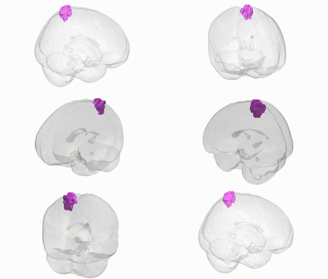
Full Quality Version: Download MP4
Wali Sidiqyar*, Gaurav Rudravaram*, Elyssa M. McMaster, Trent M. Schwartz, Adam M. Saunders, Kurt G. Schilling, Bennett A. Landman "Introducing SPINS: A Shared Public Visualization Library of Neuroanatomical Structures." Medical Imaging with Deep Learning- short paper
This resource is licensed under CC0 1.0 Universal (Public Domain).