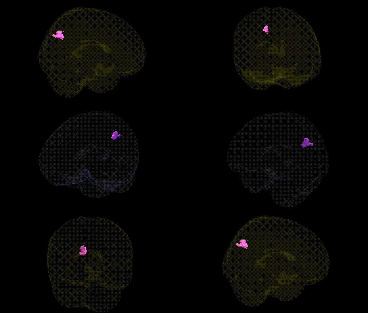
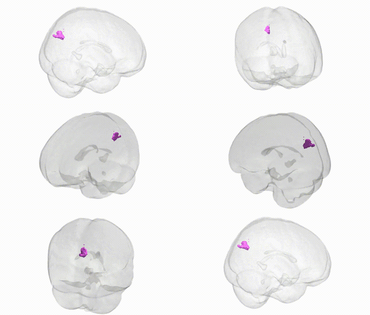
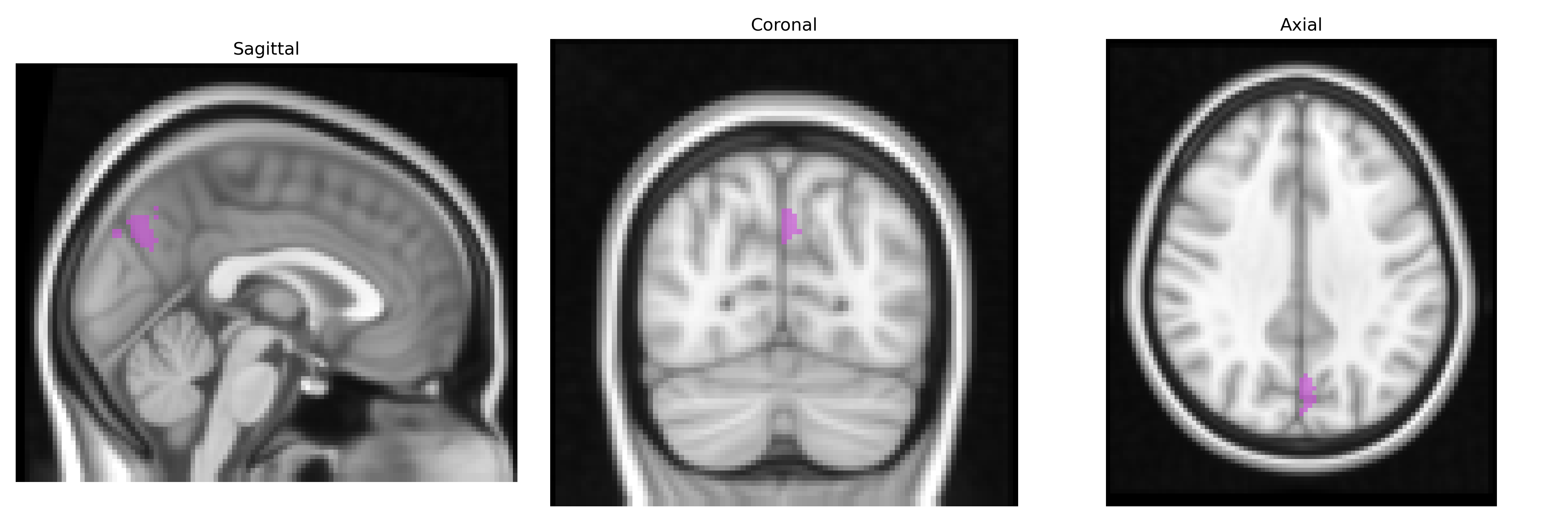
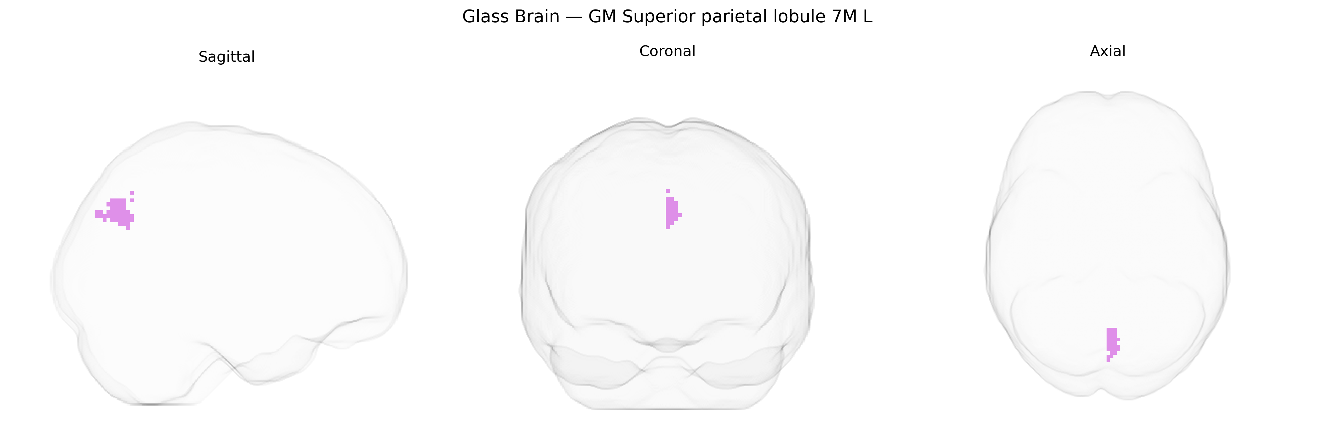
The bilateral GM Superior parietal lobule 7M L, as defined in the Juelich maxprob thr25 2mm atlas, corresponds to a cytoarchitectonic subdivision of the medial portion of the superior parietal lobule within Brodmann area 7. This region lies on the dorsal medial surface of the parietal lobe, posterior to the primary somatosensory cortex and anterior to the precuneus, and is implicated in higher-order somatosensory integration, visuospatial processing, and the coordination of attention and sensorimotor transformation. Functionally, area 7M contributes to the integration of visual and proprioceptive information for the guidance of limb and eye movements and participates in networks supporting spatial cognition and body schema representation. There is no direct Wikipedia article for “area 7M”; a closely related structure is the Superior parietal lobule.
The bilateral GM Superior parietal lobule 7M L, a medial segment of the superior parietal lobule implicated in visuospatial integration, attention, and sensorimotor coordination, shows convergent genetic associations largely through imaging genetics and GWAS of cortical structure and function rather than region-specific candidate genes. Large-scale brain morphology GWAS (e.g., ENIGMA, UK Biobank) have linked common variants in genes such as HMGA2, IGF2BP1, and various loci near microtubule- and synapse-related genes to parietal cortical thickness, surface area, and gyrification that encompass or include superior parietal regions, with polygenic architectures overlapping general cognitive ability, educational attainment, and intracranial volume. Functional imaging genetics studies implicate parietal 7M in frontoparietal and dorsal attention networks, where variants in dopamine-related genes (e.g., COMT), glutamatergic genes, and synaptic plasticity genes influence activation patterns during working memory, visuospatial tasks, and attention-demanding paradigms. Disorder-related GWAS do not typically isolate 7M specifically, but parietal association cortex, including superior parietal subdivisions, is consistently implicated in schizophrenia, ADHD, autism spectrum disorder, and Alzheimer’s disease via polygenic risk associations with parietal cortical thickness, connectivity, and default mode/frontoparietal network alterations, often mediated by genes involved in neurodevelopment, synaptic signaling, and immune pathways (e.g., MHC region, complement genes). Overall, genetic findings point to a broadly polygenic influence on structural and functional variation in the superior parietal lobule, with shared genetic architectures spanning cognition, neurodevelopmental and psychiatric disorders, and neurodegeneration, but with no single gene or variant uniquely or definitively assigned to the Juelich 7M L parcellation at current resolutions.
Overview generated by GPT-4o (2026).
Region ID: 75
Hemisphere: bilateral
Atlas: Juelich maxprob thr25 2mm
Full Quality Version: Download MP4
Full Quality Version: Download MP4
Wali Sidiqyar*, Gaurav Rudravaram*, Elyssa M. McMaster, Trent M. Schwartz, Adam M. Saunders, Kurt G. Schilling, Bennett A. Landman "Introducing SPINS: A Shared Public Visualization Library of Neuroanatomical Structures." Medical Imaging with Deep Learning- short paper
This resource is licensed under CC0 1.0 Universal (Public Domain).