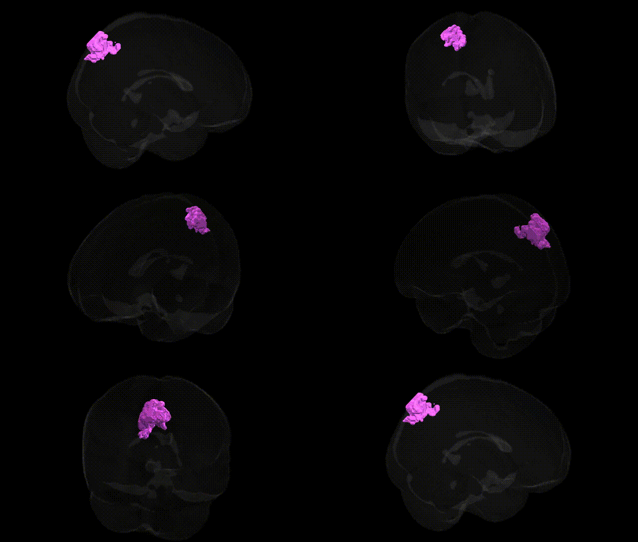
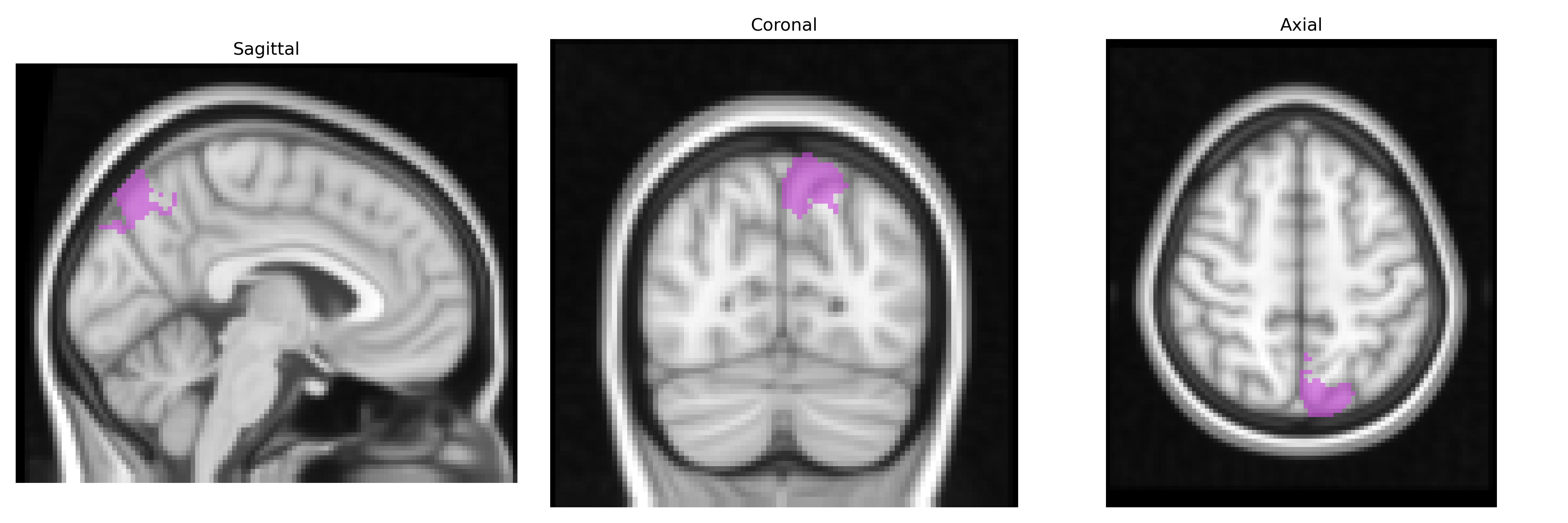
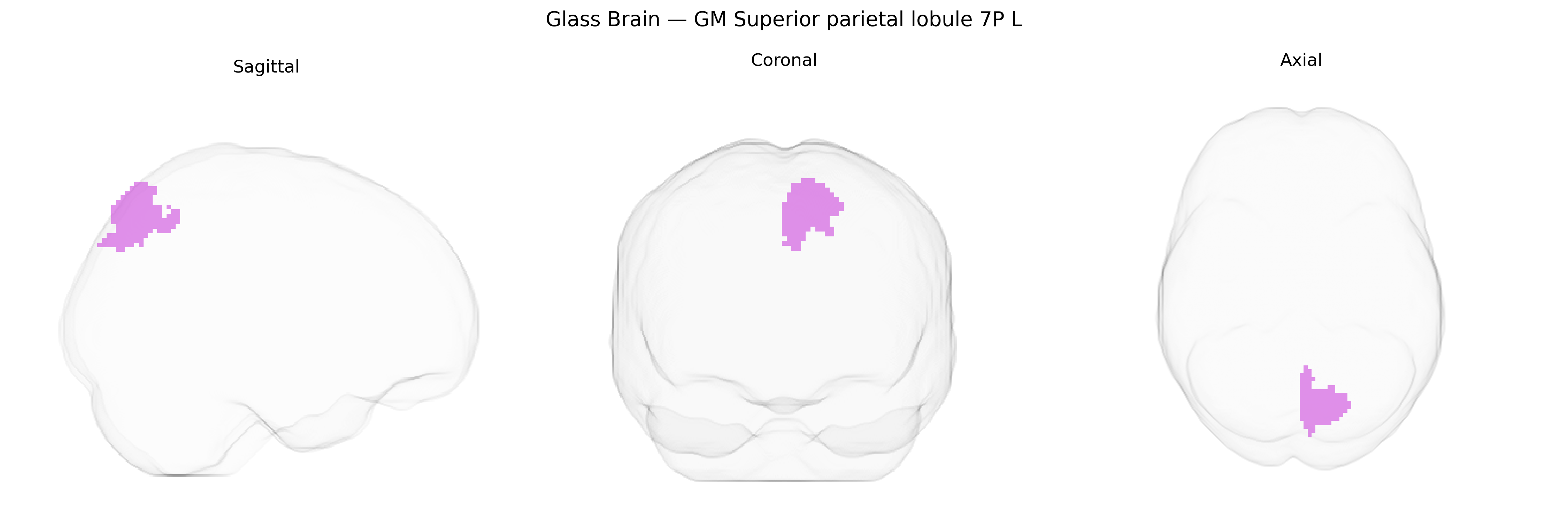
The bilateral GM Superior parietal lobule 7P L, as defined in the Juelich maxprob thr25 2mm atlas, corresponds to a cytoarchitectonic subregion (area 7P) within the superior parietal lobule of the left hemisphere, part of Brodmann area 7. This region is located on the dorsal aspect of the parietal lobe, posterior to the primary somatosensory cortex, and is implicated in higher-order somatosensory processing, visuospatial integration, and the coordination of attention and sensorimotor transformations, particularly for guiding complex, visually guided movements and spatial cognition. Cytoarchitectonically, area 7P shows characteristic granular cortical layering typical of association cortex and forms part of the posterior parietal network that interfaces sensory inputs with motor planning and executive functions. There is no direct Wikipedia article for area 7P; a related structure is the Superior parietal lobule.
The bilateral gray matter Superior Parietal Lobule area 7P (left hemisphere) from the Juelich maxprob thr25 2mm atlas corresponds to a higher-order associative parietal region implicated in visuospatial attention, sensorimotor integration, and aspects of working memory and executive control, and genetic associations with this region are typically inferred from imaging genetics and GWAS of brain structure or function rather than region-specific candidate gene studies. Large-scale GWAS of cortical thickness and surface area (e.g., ENIGMA and UK Biobank cohorts) have identified multiple loci (including variants near genes such as KIAA0586, CENPW, DACT1, and others involved in neurodevelopment and synaptic function) that influence parietal cortical morphology, with several signals mapping to superior parietal parcels or broader parietal territories that encompass 7P. Polygenic risk scores for schizophrenia, major depressive disorder, bipolar disorder, and attention-deficit/hyperactivity disorder have been associated with altered parietal volume or functional activation, and functional imaging genetics studies link common variants in dopaminergic, glutamatergic, and neurodevelopmental genes (e.g., COMT Val158Met, BDNF Val66Met, DISC1, CACNA1C) to variability in superior parietal activation during tasks requiring attention shifting, visuospatial working memory, and motor planning. In addition, GWAS of cognitive traits—such as general cognitive ability, processing speed, mathematical performance, and educational attainment—frequently report parietal structural and functional correlates, suggesting that polygenic influences on cognition act partly through superior parietal networks that include 7P. Neurodevelopmental disorders with strong genetic components (autism spectrum conditions, dyslexia, developmental coordination disorder) and neurodegenerative or cerebrovascular conditions (Alzheimer’s disease, small vessel disease) also show reproducible involvement of superior parietal regions, and imaging–genetic correlation analyses indicate that risk variants in synaptic, axonal guidance, and immune-related genes affect parietal morphology and connectivity. However, current evidence is largely parcel- or network-level rather than specific to the Juelich 7P L definition, and no single gene or variant is uniquely or exclusively associated with this precise cytoarchitectonic region; instead, a distributed set of common and rare variants influencing cortical development, synaptic plasticity, and white-matter connectivity contribute probabilistically to structural and functional variation in the superior parietal lobule, including area 7P.
Overview generated by GPT-4o (2026).
Region ID: 79
Hemisphere: bilateral
Atlas: Juelich maxprob thr25 2mm
Full Quality Version: Download MP4
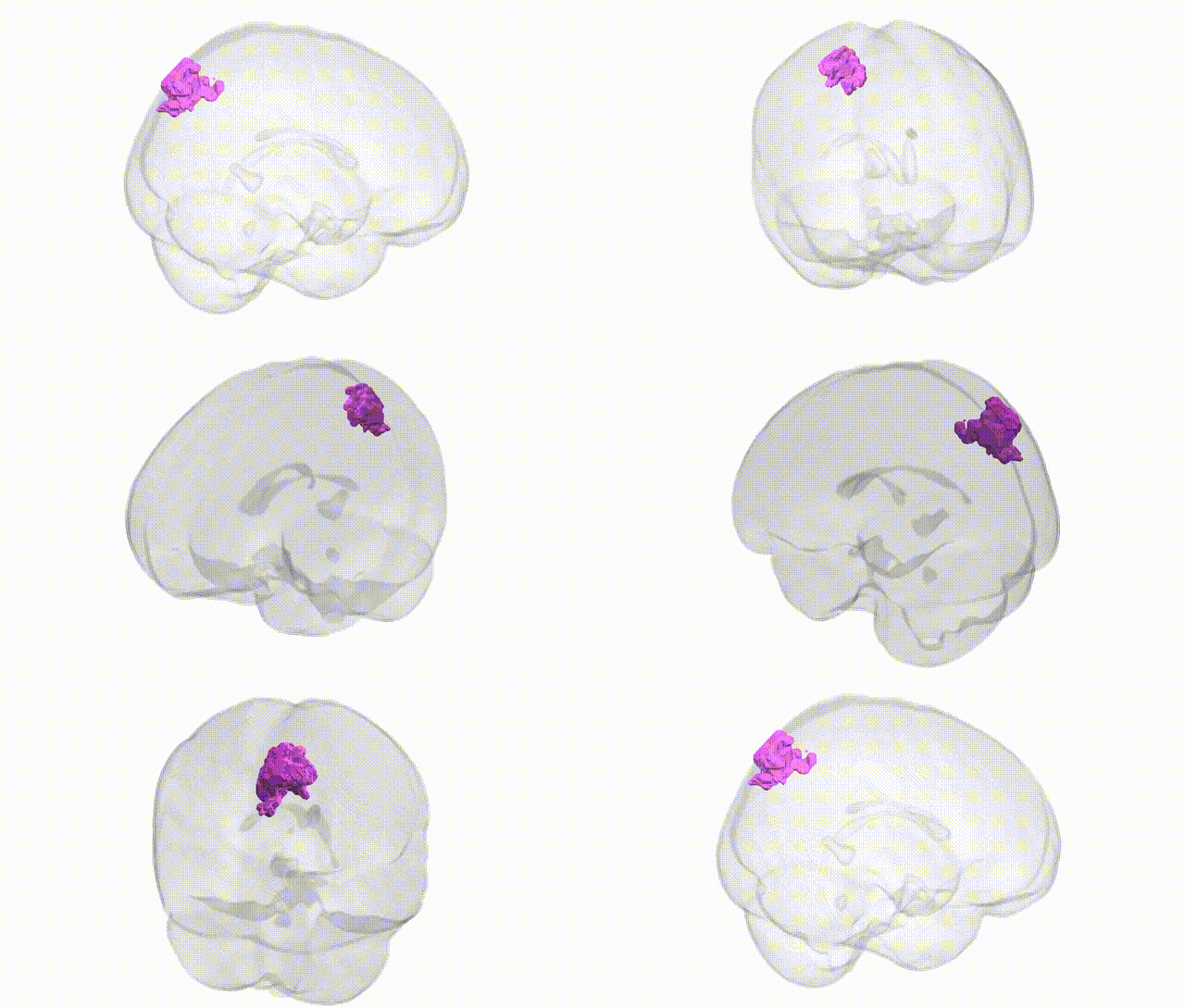
Full Quality Version: Download MP4
Wali Sidiqyar*, Gaurav Rudravaram*, Elyssa M. McMaster, Trent M. Schwartz, Adam M. Saunders, Kurt G. Schilling, Bennett A. Landman "Introducing SPINS: A Shared Public Visualization Library of Neuroanatomical Structures." Medical Imaging with Deep Learning- short paper
This resource is licensed under CC0 1.0 Universal (Public Domain).