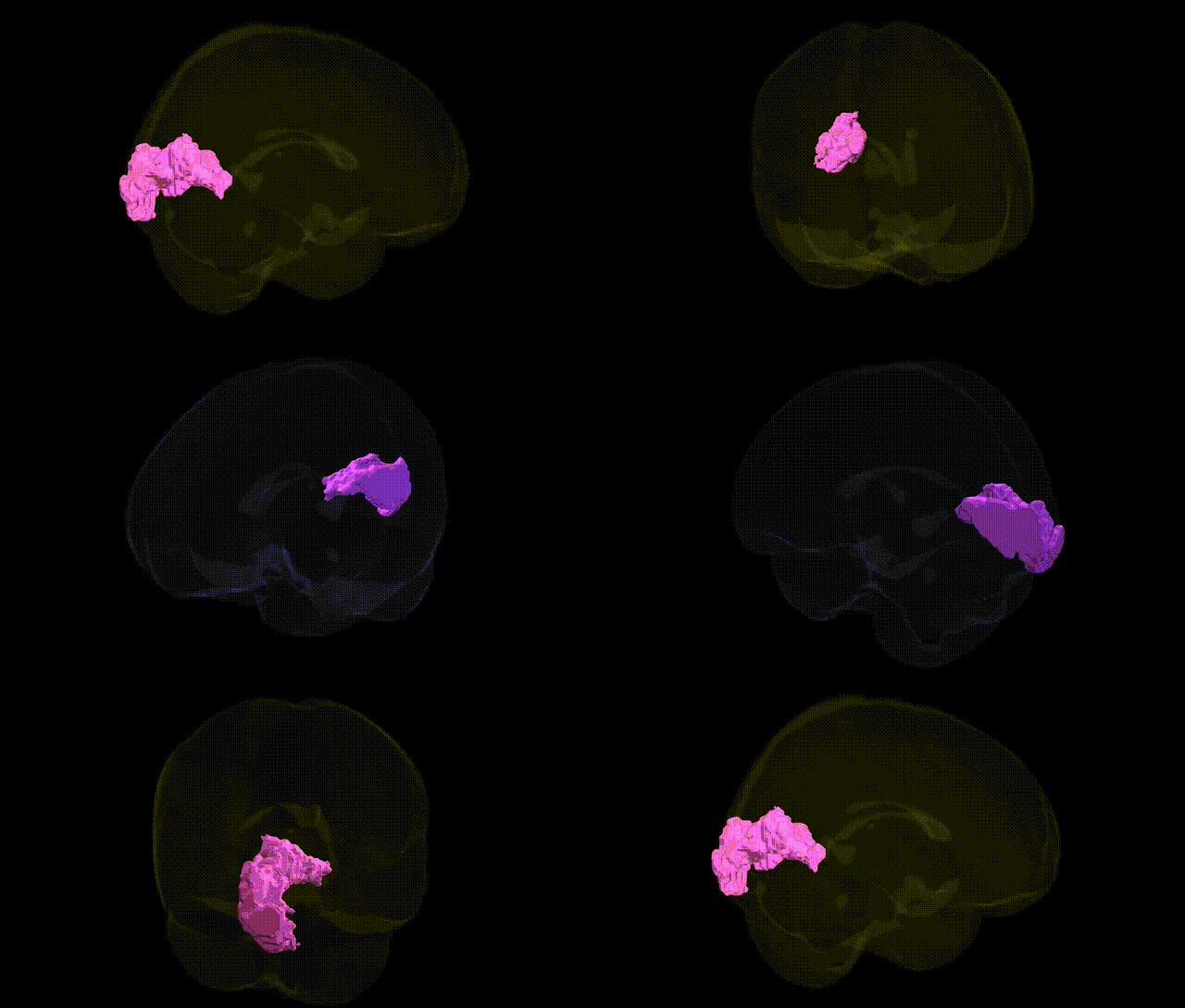
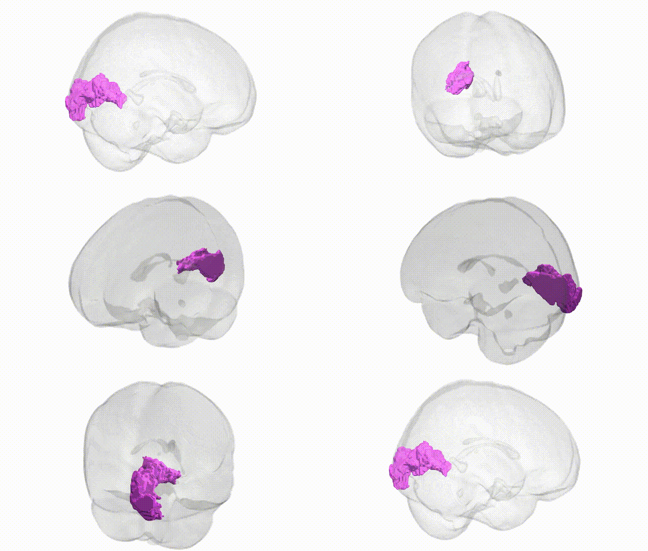
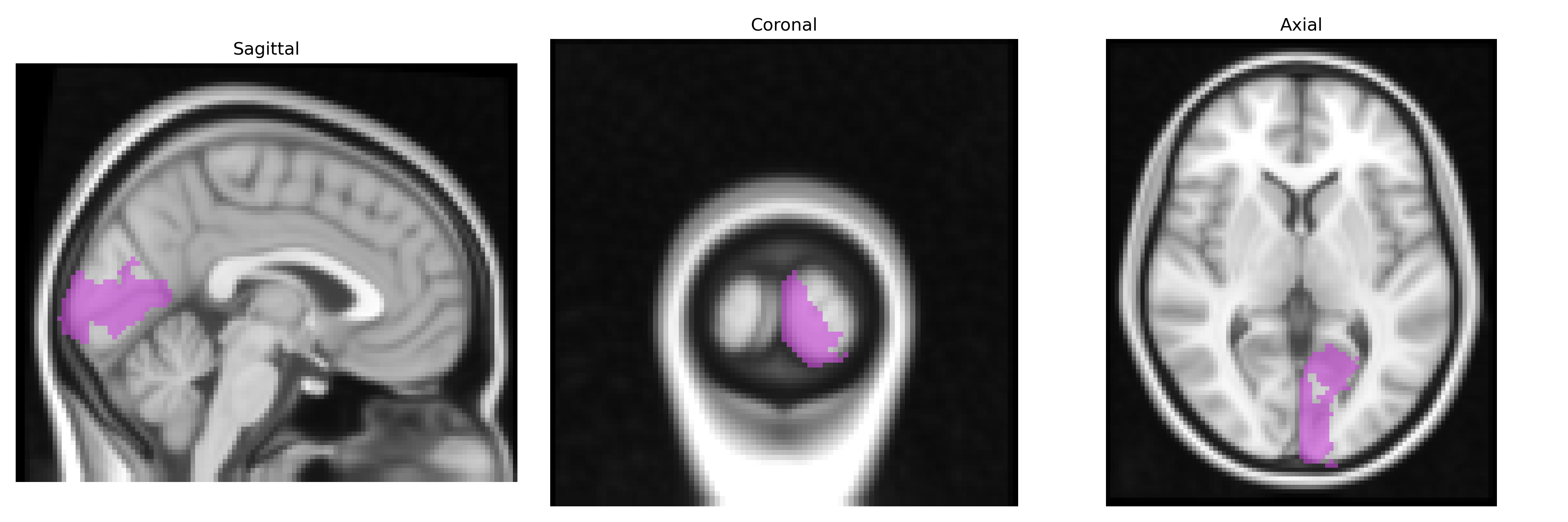
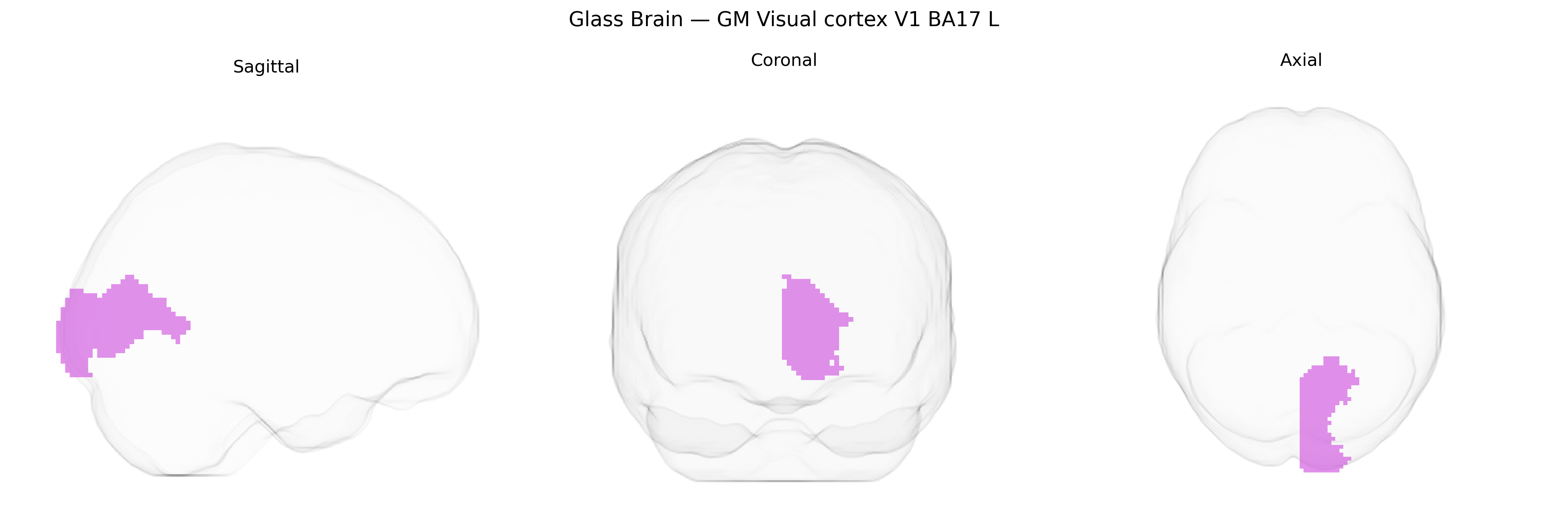
The bilateral GM Visual cortex V1 BA17 L region in the Juelich maxprob thr25 2 mm Atlas corresponds to the left primary visual cortex (V1), cytoarchitectonically defined as Brodmann area 17, located along the banks of the calcarine sulcus in the occipital lobe. This cortical area contains a precise retinotopic map of the visual field and represents the earliest stage of cortical visual processing, receiving its main afferent input from the lateral geniculate nucleus of the thalamus via the optic radiation. Neurons in this region are highly sensitive to basic visual features such as orientation, spatial frequency, contrast, and binocular disparity, providing the foundational representations that are further processed by higher-order visual areas (e.g., V2, V3) for form, motion, and color perception. Although the atlas label specifies the left hemisphere, V1 is present bilaterally and exhibits strong interhemispheric homology in structure and function. Primary visual cortex
The bilateral primary visual cortex (V1, Brodmann area 17, as defined in the Juelich maxprob thr25 2 mm atlas) shows heritable variation in cortical thickness, surface area, and volume, with SNP-based heritability estimates from large imaging–genetics consortia such as ENIGMA and UK Biobank typically in the 20–40% range for structural metrics. Genome-wide association studies of occipital and specifically V1-related measures have implicated common variants in or near genes involved in neurodevelopment, axon guidance, synaptic plasticity, and myelination (for example, loci near genes such as MAPT, WNT and FGF pathway components, and other regulators of cortical patterning), although associations are often shared across multiple visual or occipital regions rather than unique to V1. Polygenic architectures overlapping with general cognitive ability, educational attainment, and intracranial volume have been reported, suggesting that some variants influencing global brain development and cortical scaling also affect V1 structure. Genetic links between V1 and neuropsychiatric disorders are indirect: schizophrenia, bipolar disorder, autism spectrum disorder, and major depression show polygenic overlap with global and regional cortical measures, including occipital cortex, but no single disorder-defining locus has been uniquely assigned to V1. In contrast, primary visual phenotypes such as refractive error, visual acuity, and amblyopia have distinct GWAS signals largely in ocular or anterior visual pathway genes rather than cortex-specific loci, with downstream effects on V1 considered more environmentally or activity driven. Overall, current evidence supports moderate heritability and polygenic influences on V1 structure and function, with shared genetic architectures spanning broader visual and association cortex rather than V1-specific disease loci.
Overview generated by GPT-4o (2026).
Region ID: 81
Hemisphere: bilateral
Atlas: Juelich maxprob thr25 2mm
Full Quality Version: Download MP4
Full Quality Version: Download MP4
Wali Sidiqyar*, Gaurav Rudravaram*, Elyssa M. McMaster, Trent M. Schwartz, Adam M. Saunders, Kurt G. Schilling, Bennett A. Landman "Introducing SPINS: A Shared Public Visualization Library of Neuroanatomical Structures." Medical Imaging with Deep Learning- short paper
This resource is licensed under CC0 1.0 Universal (Public Domain).