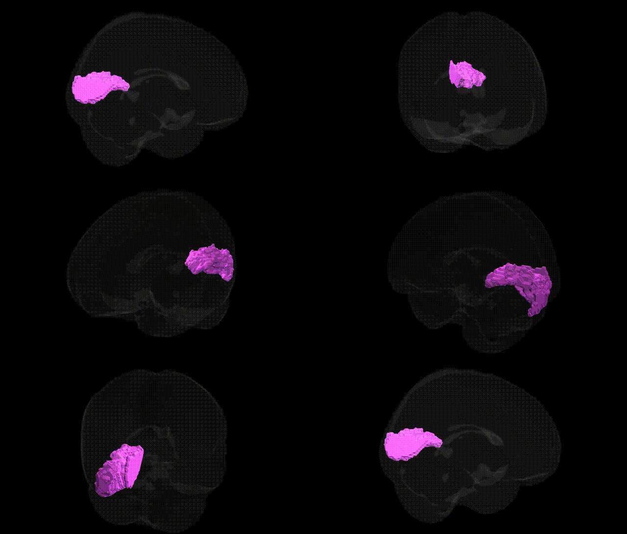
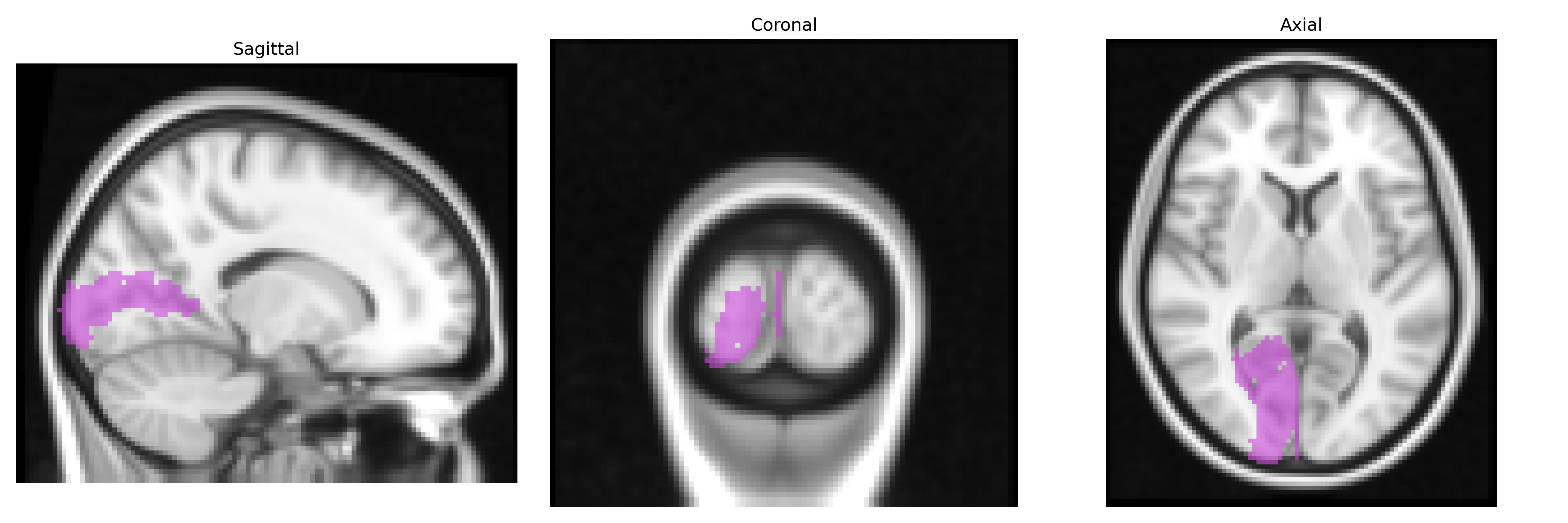
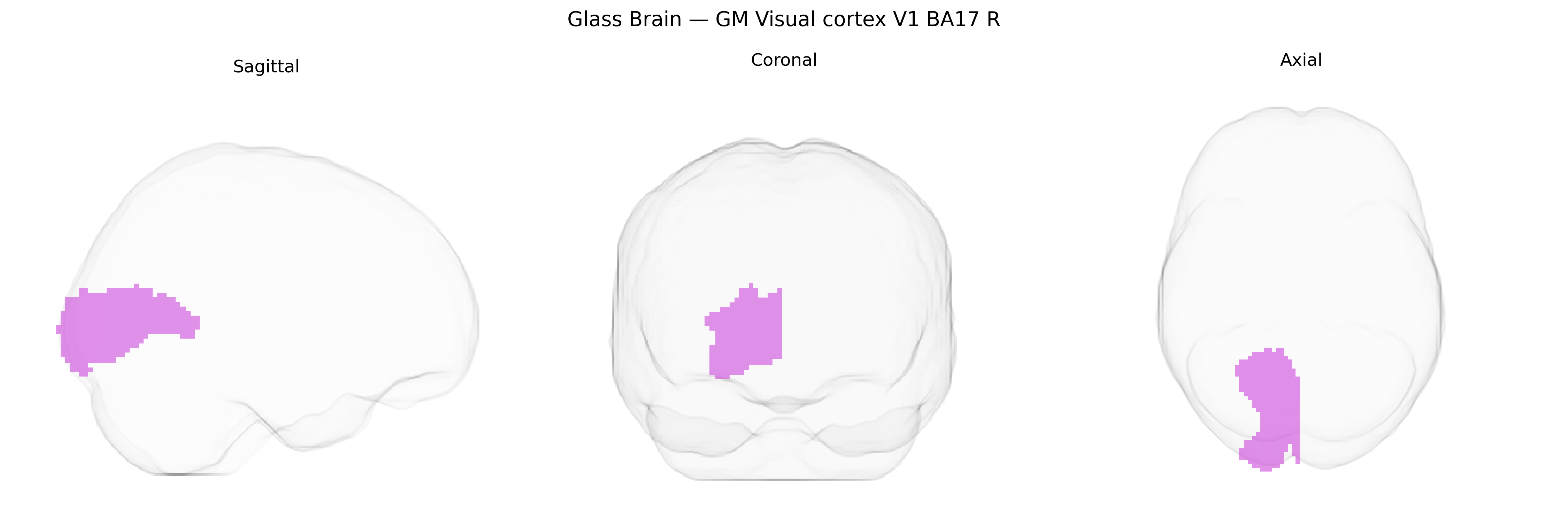
The bilateral GM Visual cortex V1 BA17 R region from the Juelich maxprob thr25 2mm atlas corresponds to the right primary visual cortex (V1), located in the medial occipital lobe along the banks of the calcarine sulcus and representing the earliest cortical stage of visual processing. This region is cytoarchitectonically defined as Brodmann area 17 and is characterized by a highly laminated structure, including a prominent granular layer IV that receives dense thalamocortical input from the lateral geniculate nucleus. Functionally, it encodes fundamental attributes of the visual scene such as orientation, spatial frequency, contrast, and basic motion, and exhibits a precise retinotopic organization with foveal input represented posteriorly and peripheral visual fields represented more anteriorly along the calcarine fissure. The “R” designation indicates right-hemispheric localization, but the atlas label is part of a bilateral group capturing gray matter voxels most likely assigned to V1 at a 25% probability threshold. Primary visual cortex
The bilateral GM visual cortex V1 (BA17, right hemisphere) from the Juelich maxprob thr25 2 mm atlas corresponds to primary visual cortex, a region whose structure and function show substantial heritability and multiple genetic associations from imaging-genetics and GWAS studies. Large-scale brain MRI GWAS (e.g., ENIGMA, UK Biobank–based analyses) have identified variants in genes involved in neurodevelopment, axon guidance, synaptic function, and myelination—such as NRXN1, PAX6, TCF4, and others—associated with occipital and primary visual cortex surface area, thickness, or volume, often as part of broader cortical measures. Polygenic risk for neuropsychiatric disorders including schizophrenia, bipolar disorder, major depressive disorder, and autism spectrum disorder has been linked to structural and functional alterations in V1/occipital cortex, with some studies showing that schizophrenia- and ASD-associated loci relate to reduced visual cortex thickness or altered connectivity. Visual cortex traits, including cortical folding patterns and surface area, have also been tied to common variants in genes affecting ocular and visual system development (for example, loci near GDF6 and other eye-development genes), while genetic risk for neurodegenerative diseases such as Alzheimer’s disease and Parkinson’s disease has been associated with occipital/visual cortex atrophy patterns in some imaging-genetic analyses. Although many findings are not specific to the right BA17 subregion but rather to bilateral or global occipital/visual cortex measures, converging evidence supports that primary visual cortex morphology and function are influenced by a polygenic architecture overlapping with genes implicated in neurodevelopment, psychiatric risk, visual system development, and general brain structure.
Overview generated by GPT-4o (2026).
Region ID: 82
Hemisphere: bilateral
Atlas: Juelich maxprob thr25 2mm
Full Quality Version: Download MP4
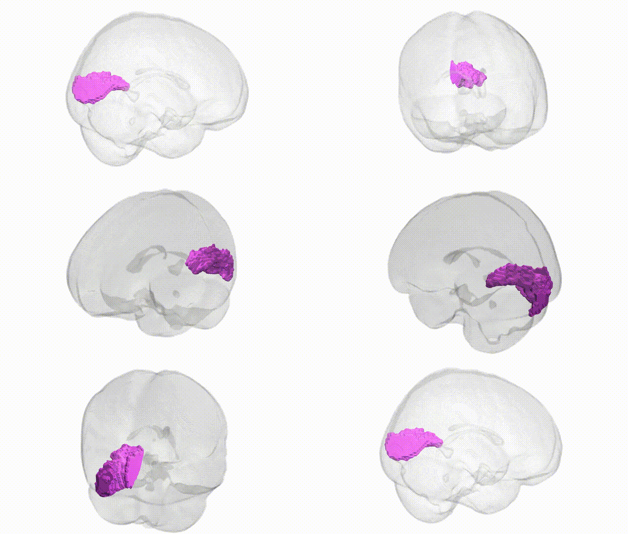
Full Quality Version: Download MP4
Wali Sidiqyar*, Gaurav Rudravaram*, Elyssa M. McMaster, Trent M. Schwartz, Adam M. Saunders, Kurt G. Schilling, Bennett A. Landman "Introducing SPINS: A Shared Public Visualization Library of Neuroanatomical Structures." Medical Imaging with Deep Learning- short paper
This resource is licensed under CC0 1.0 Universal (Public Domain).