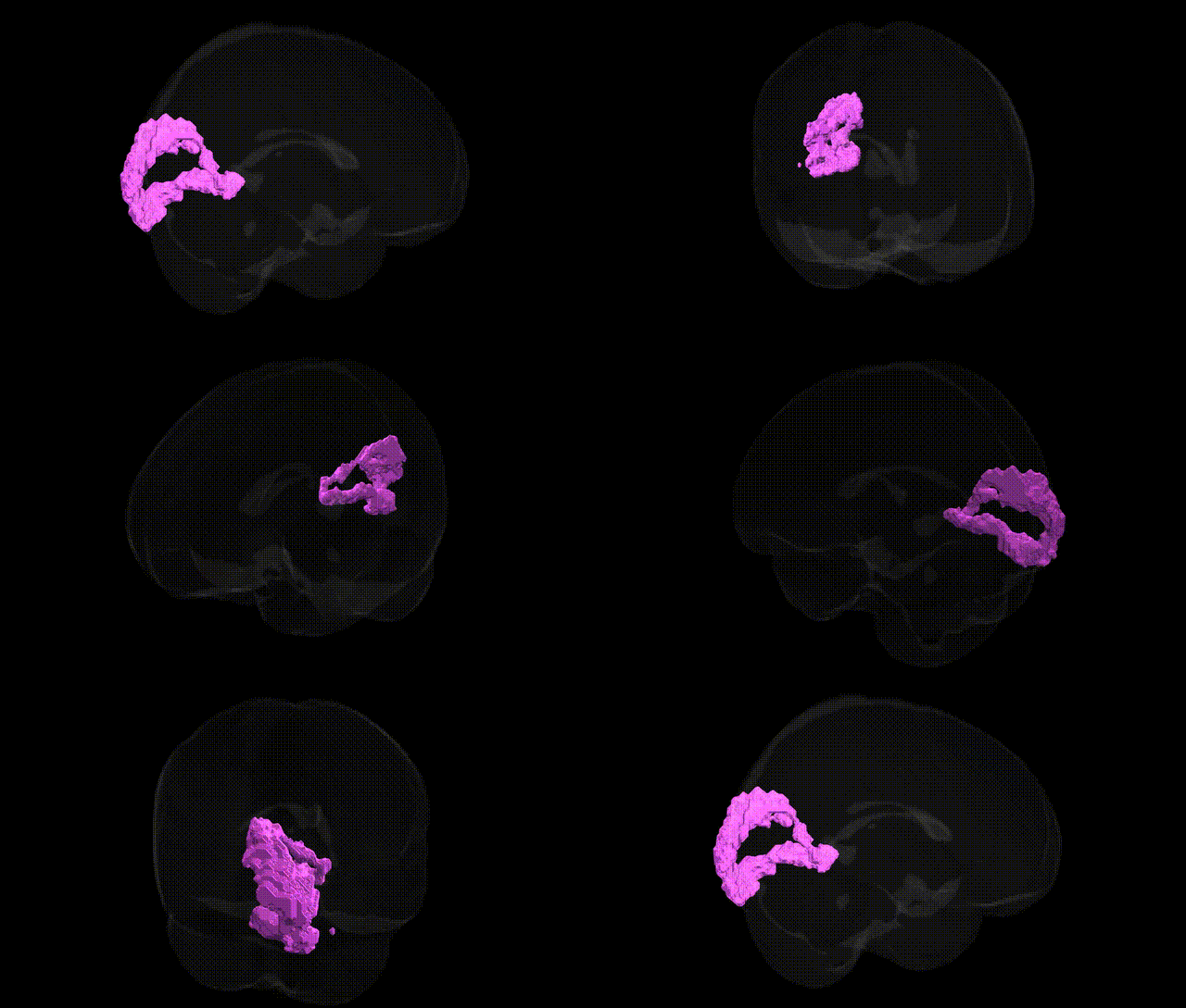
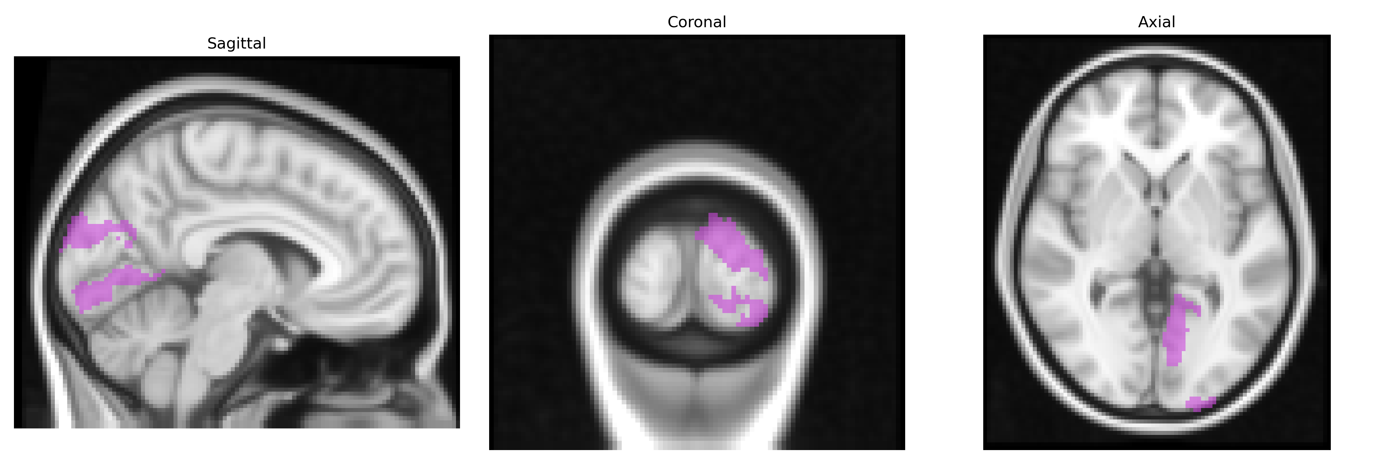
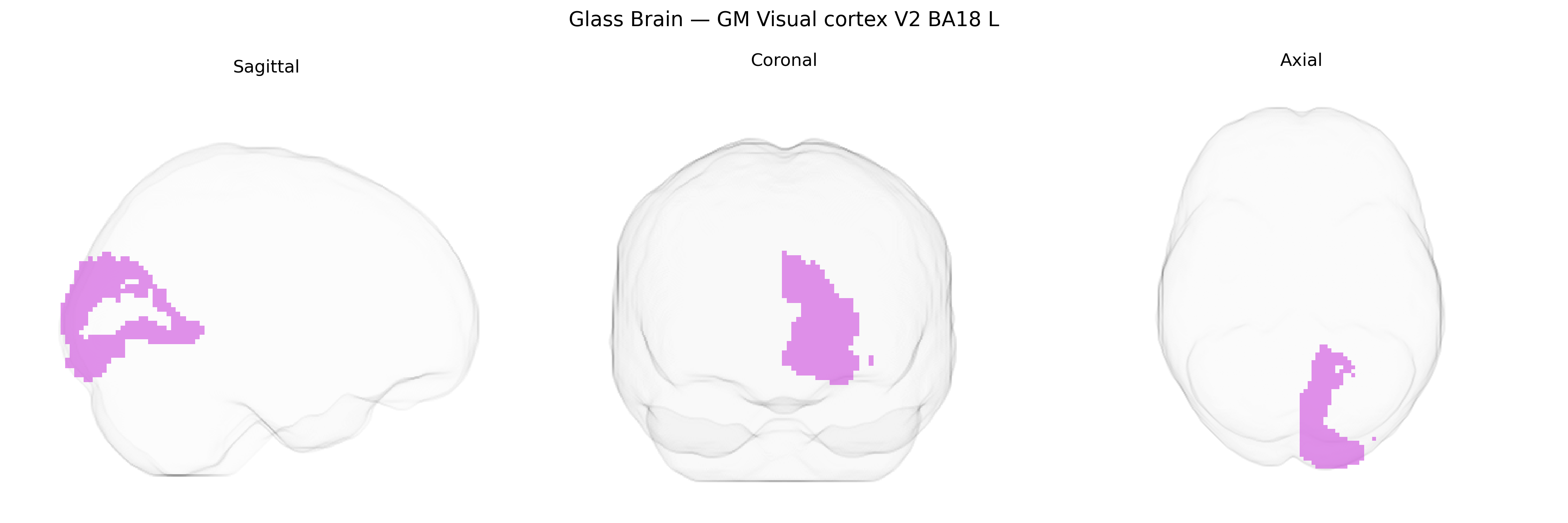
The bilateral GM Visual cortex V2 BA18 L region in the Juelich maxprob thr25 2mm atlas corresponds to the left-hemisphere portion of secondary visual cortex (area V2), largely overlapping cytoarchitectonic Brodmann area 18. It lies adjacent to primary visual cortex (V1/BA17) along the banks of the calcarine sulcus in the occipital lobe and receives dense feedforward input from V1 while projecting to higher-order visual areas (e.g., V3, V4, MT). Neuronal populations in V2 are organized in modular patterns that process intermediate-level visual features, including orientation, spatial frequency, contours, binocular disparity, and simple form properties, serving as a crucial stage for transforming retinotopic signals into more complex representations used in object recognition and visuospatial analysis. There is no direct link for “Visual cortex V2 BA18”; see the related structure Visual area V2.
The bilateral grey matter visual cortex V2 (Brodmann area 18, left hemisphere) from the Juelich maxprob thr25 2 mm atlas has been implicated in genetic studies of visual processing, cortical structure, and neuropsychiatric risk, although most findings concern broader occipital or visual association regions rather than BA18 specifically. GWAS of cortical thickness and surface area have identified multiple loci (including variants near genes such as MAPT, KIAA0586, PAX6, and others involved in neurodevelopment and synaptic function) associated with occipital and visual cortical morphology that encompass or overlap V2/BA18, suggesting polygenic influences on the structural integrity and organization of this region. Genetic studies of visual perception, including contrast sensitivity and visual acuity, have linked variation in visual cortex function to genes involved in myelination and neurotransmission, while imaging genetics work in disorders such as schizophrenia, major depression, and autism has reported risk alleles (for example in CACNA1C, GRIN2A, and complement pathway genes) associated with altered activation or connectivity in secondary visual areas that include V2. In migraine (especially with visual aura), GWAS hits in genes modulating cortical excitability and vascular regulation have been connected to altered visual cortex responsiveness, and in neurodegenerative disease, occipital/visual cortex atrophy patterns show associations with APOE and other Alzheimer’s disease risk loci, indirectly implicating BA18 in genetically mediated vulnerability of posterior cortical networks. Overall, existing evidence points to a distributed, polygenic architecture in which susceptibility genes for brain structure, visual perception, and several neuropsychiatric and neurological disorders influence the morphology and functional responsivity of the V2/BA18 region, though highly specific gene–BA18 associations remain limited and are usually inferred from broader occipital or visual-system analyses.
Overview generated by GPT-4o (2026).
Region ID: 83
Hemisphere: bilateral
Atlas: Juelich maxprob thr25 2mm
Full Quality Version: Download MP4
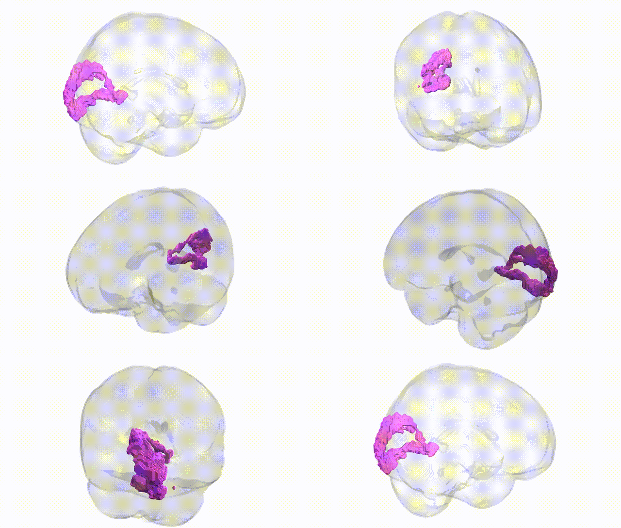
Full Quality Version: Download MP4
Wali Sidiqyar*, Gaurav Rudravaram*, Elyssa M. McMaster, Trent M. Schwartz, Adam M. Saunders, Kurt G. Schilling, Bennett A. Landman "Introducing SPINS: A Shared Public Visualization Library of Neuroanatomical Structures." Medical Imaging with Deep Learning- short paper
This resource is licensed under CC0 1.0 Universal (Public Domain).