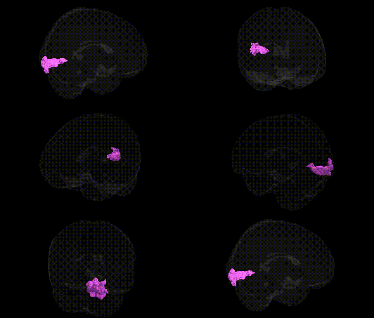
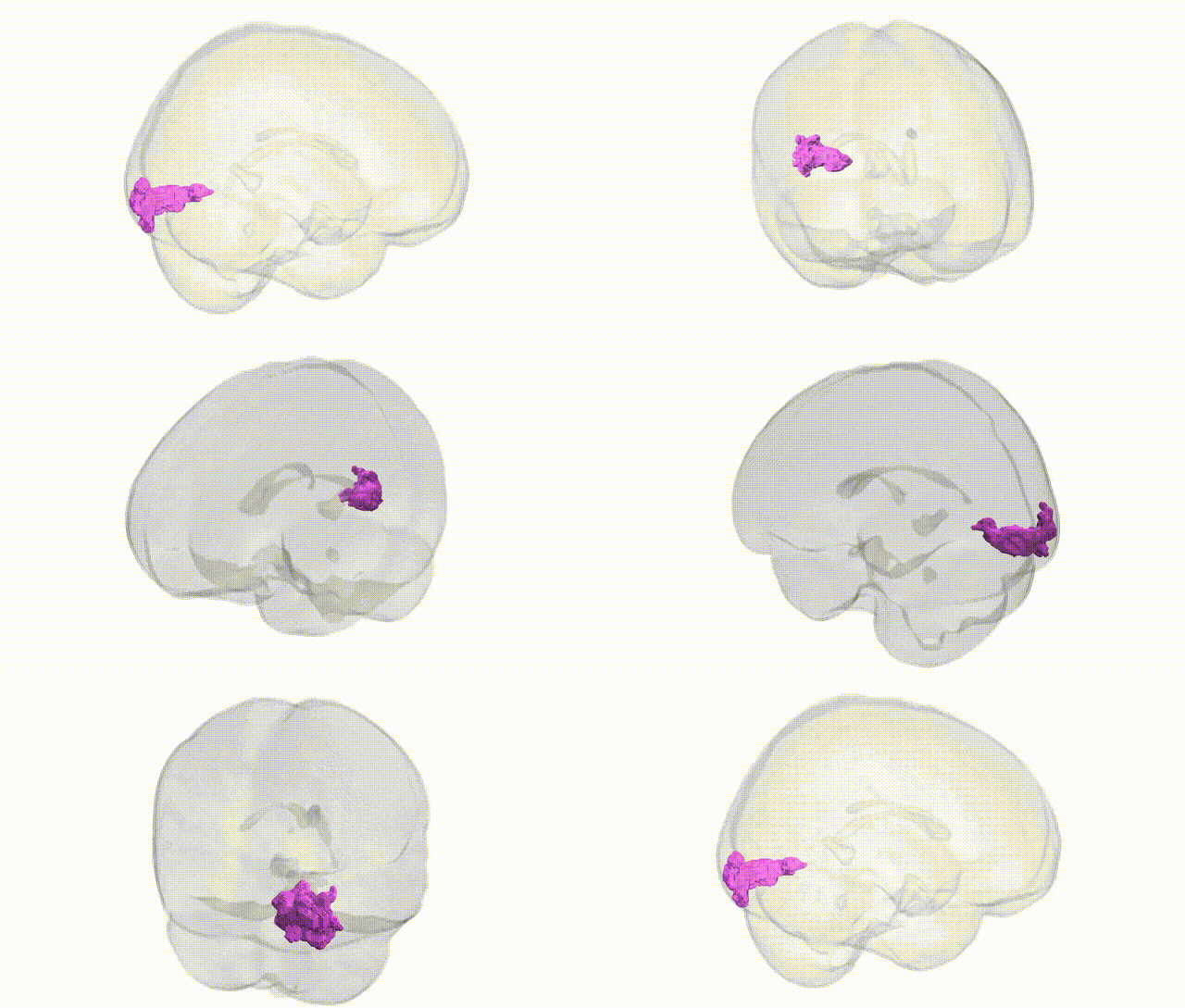
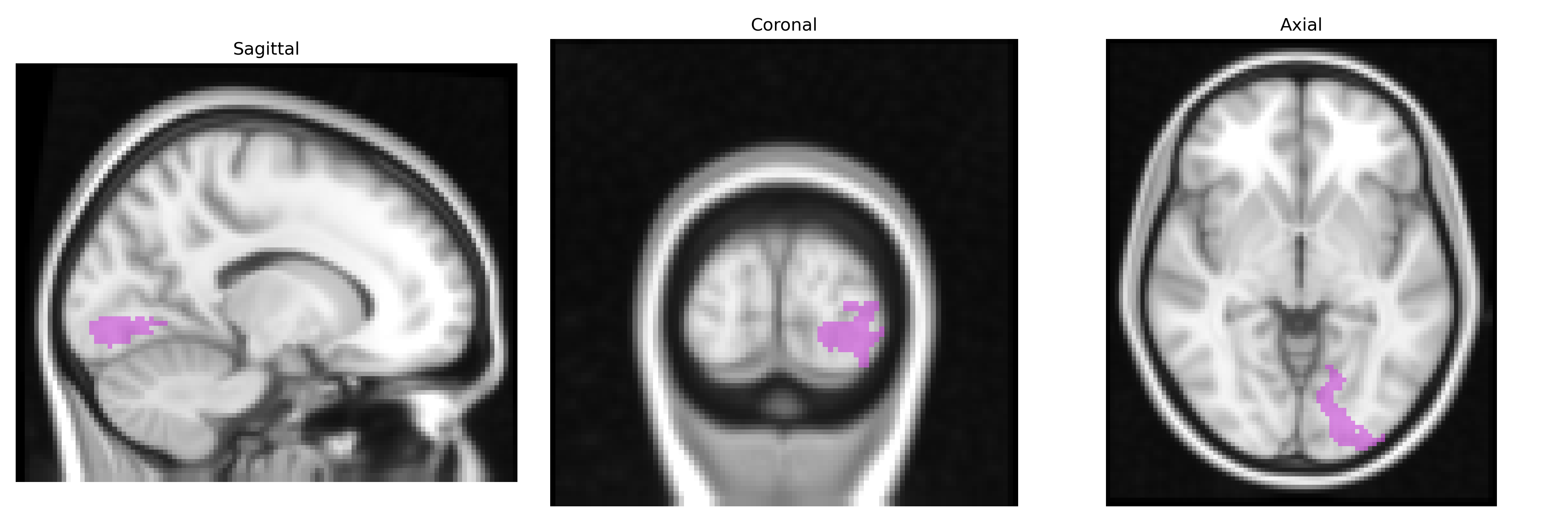
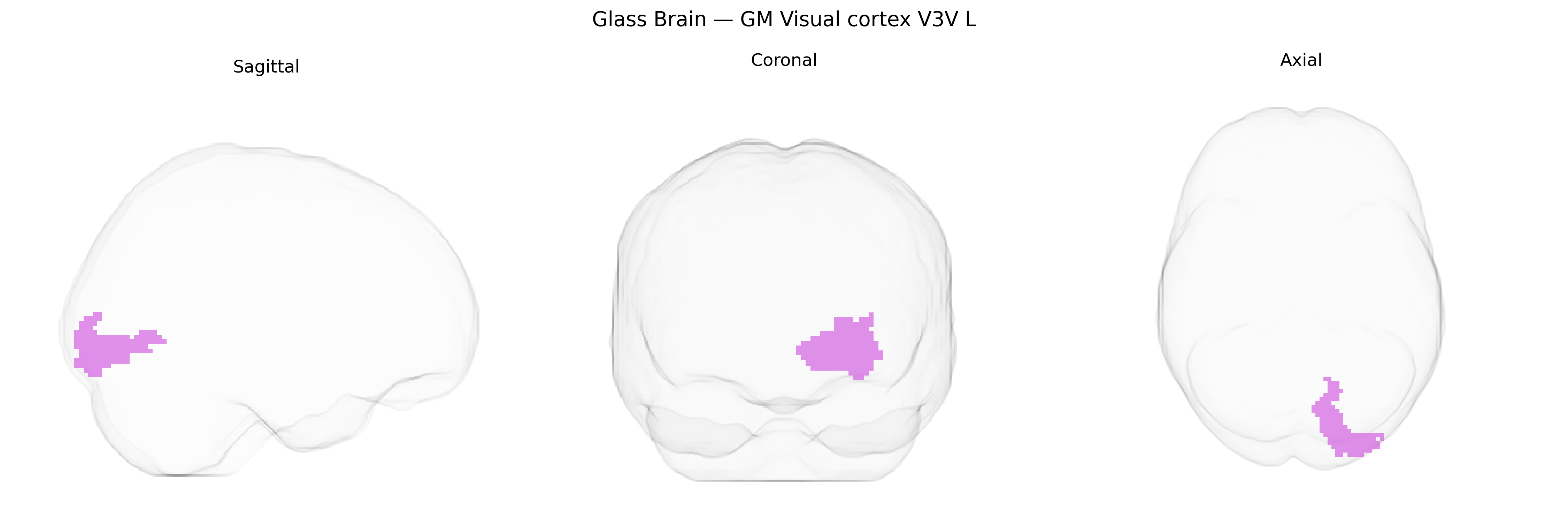
The bilateral GM Visual cortex V3V L region, as defined in the Juelich maxprob thr25 2mm Atlas, corresponds to the ventral portion of visual area V3 in the left hemisphere, located in the occipital lobe along the ventral bank of the calcarine sulcus and extending into adjacent occipitotemporal cortex. This region is part of the early extrastriate visual cortex and participates in the processing of visual information related to form, orientation, and motion, with a bias toward the lower visual field representation. V3V receives strong feedforward input from primary visual cortex (V1) and V2 and sends projections to higher-order ventral stream areas such as V4 and inferotemporal cortex, integrating local visual features into more complex representations that support object recognition and scene analysis. There is no direct Wikipedia article for V3V; a closely related and encompassing structure is the visual cortex: Visual cortex.
The bilateral GM Visual cortex V3V L region, as defined in the Juelich maxprob thr25 2mm atlas, lies within extrastriate visual cortex implicated in intermediate-level visual processing, and genetic associations involving this area generally emerge from imaging genomics and GWAS of brain structure and function rather than region-specific candidate gene studies. Large-scale GWAS of cortical thickness and surface area have identified variants in neurodevelopmental and synaptic genes (e.g., MAPT on 17q21.31, genes in Wnt signaling and axon guidance pathways) that influence occipital and visual cortical morphology, with some effects overlapping regions encompassing V3/V3v, and these structural measures have been genetically correlated with cognitive traits, educational attainment, and general intelligence. Polygenic risk scores for neuropsychiatric disorders such as schizophrenia, bipolar disorder, and major depression have been associated with altered occipital and visual cortex volume or activation, including in regions near V3, suggesting shared genetic architecture between visual cortical organization and vulnerability to these conditions. Additionally, GWAS of functional MRI measures, visual processing endophenotypes, and resting-state networks involving occipital cortex have pointed to common variants in genes related to synaptic plasticity and glutamatergic signaling, though findings typically reference broader “visual cortex” or occipital lobes rather than the V3V L parcel specifically. Overall, current genetic evidence links variation in and around this visual cortical region to neurodevelopmental pathways, individual differences in cognition and perception, and polygenic liability to psychiatric disorders, but precise, parcel-specific GWAS associations for V3V L remain limited, reflecting the resolution and conventions of existing brain–genome mapping studies.
Overview generated by GPT-4o (2026).
Region ID: 85
Hemisphere: bilateral
Atlas: Juelich maxprob thr25 2mm
Full Quality Version: Download MP4
Full Quality Version: Download MP4
Wali Sidiqyar*, Gaurav Rudravaram*, Elyssa M. McMaster, Trent M. Schwartz, Adam M. Saunders, Kurt G. Schilling, Bennett A. Landman "Introducing SPINS: A Shared Public Visualization Library of Neuroanatomical Structures." Medical Imaging with Deep Learning- short paper
This resource is licensed under CC0 1.0 Universal (Public Domain).