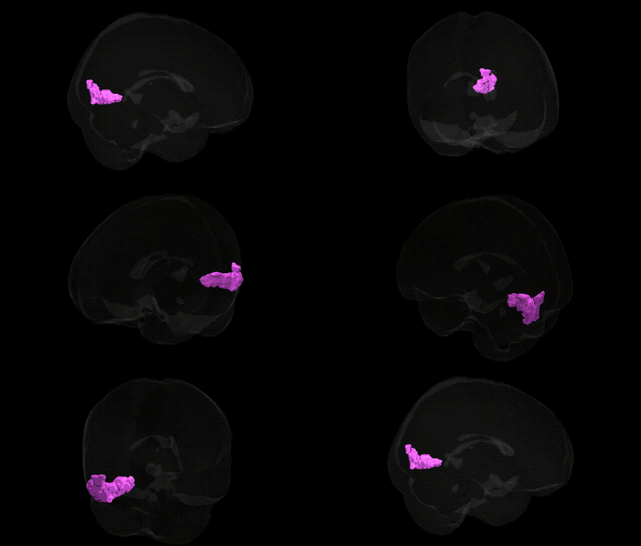
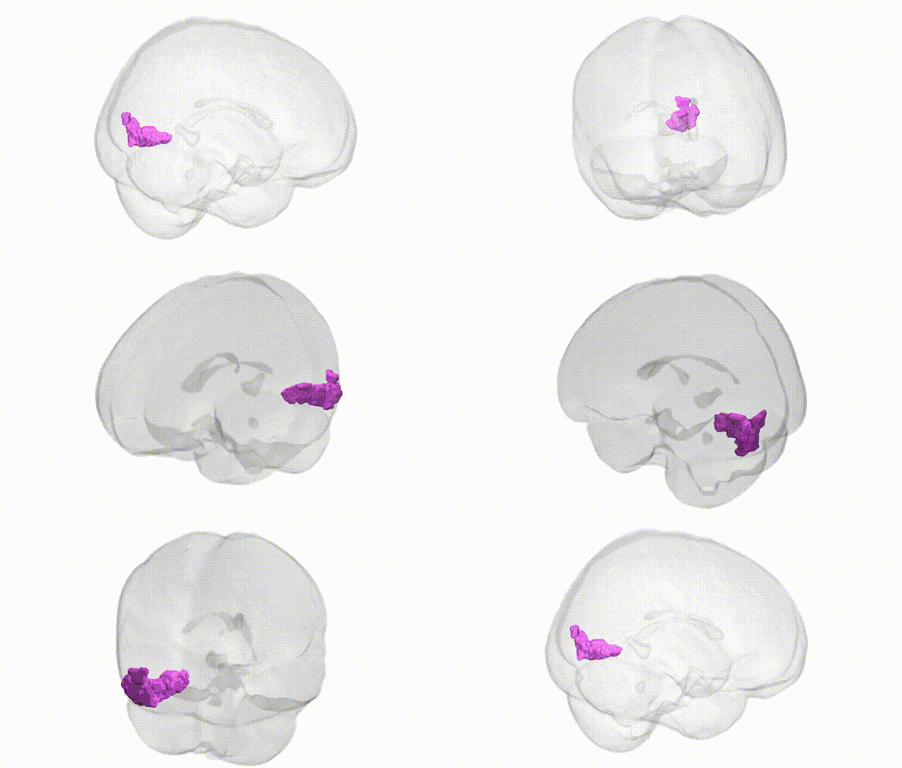
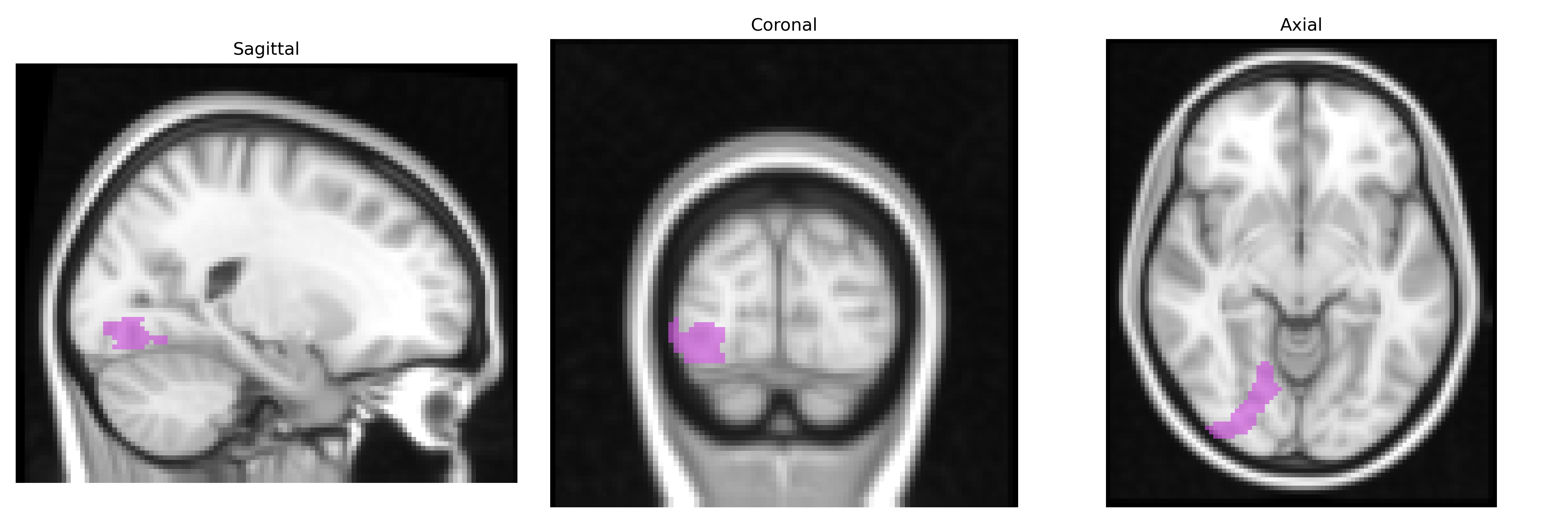
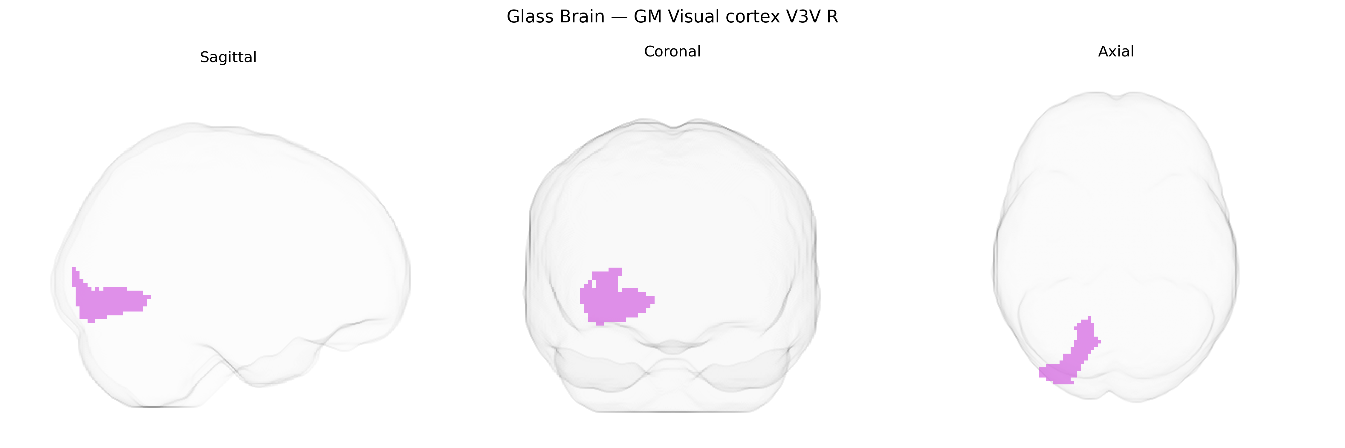
The bilateral GM Visual cortex V3V R region from the Juelich maxprob thr25 2mm Atlas corresponds to the ventral portion of visual area V3 in the right occipital lobe, forming part of the early extrastriate visual cortex. V3V is situated anterior to primary visual cortex (V1) and secondary visual cortex (V2), along the ventral occipital surface, and is implicated in processing dynamic visual information such as form, orientation, and motion, with stronger involvement in the ventral (object-related) visual stream. Cytoarchitectonically, it exhibits distinct laminar organization characteristic of association visual areas, receiving feedforward input from V1/V2 and projecting to higher-order ventral stream regions, thereby contributing to the transformation of basic visual signals into more complex perceptual representations. There is no direct Wikipedia article for V3V; a closely related structure is Visual area V3.
The bilateral gray matter visual cortex V3V (right hemisphere) in the Juelich maxprob atlas corresponds to mid-level extrastriate visual areas implicated in shape, motion, and object processing, and genetic associations typically emerge from large imaging-genetics and GWAS studies of cortical structure and functional networks rather than region-specific candidate gene work. Genome-wide analyses of cortical thickness and surface area (e.g., ENIGMA and UK Biobank consortia) have identified common variants in loci such as 15q14 (near C15orf54), 14q23.1 (KIAA0586/DAAM1 region), and genes regulating neurodevelopmental patterning (e.g., FGFRs, TBR1-related pathways) that influence occipital and visual cortical measures, with these structural variants sometimes overlapping risk loci for neurodevelopmental disorders and psychiatric conditions, including schizophrenia, autism spectrum disorder, and ADHD, which often show alterations in visual association cortices. Polygenic scores for cognitive performance, educational attainment, and general intelligence have also been associated with variability in occipital cortical thickness and functional connectivity in visual networks encompassing V3/V3V, suggesting pleiotropic effects of neurodevelopmental and synaptic genes on both visual processing architecture and higher cognitive traits. Moreover, GWAS of migraine and visual aura, as well as visual hallucinations in Parkinson’s disease and Lewy body dementia, implicate genetic pathways related to excitatory–inhibitory balance (e.g., glutamatergic and GABAergic signaling genes) that may modulate excitability in extrastriate visual regions including V3V, although these associations are typically reported at the level of broader occipital or visual networks rather than the specific V3V parcel. Overall, current genetic evidence links variation in neurodevelopmental, synaptic, and cortical patterning genes to structural and functional characteristics of occipital visual association cortex, with downstream relations to neurodevelopmental, psychiatric, and sensory phenotypes that likely involve V3V but rarely isolate it as a unique genetic target.
Overview generated by GPT-4o (2026).
Region ID: 86
Hemisphere: bilateral
Atlas: Juelich maxprob thr25 2mm
Full Quality Version: Download MP4
Full Quality Version: Download MP4
Wali Sidiqyar*, Gaurav Rudravaram*, Elyssa M. McMaster, Trent M. Schwartz, Adam M. Saunders, Kurt G. Schilling, Bennett A. Landman "Introducing SPINS: A Shared Public Visualization Library of Neuroanatomical Structures." Medical Imaging with Deep Learning- short paper
This resource is licensed under CC0 1.0 Universal (Public Domain).