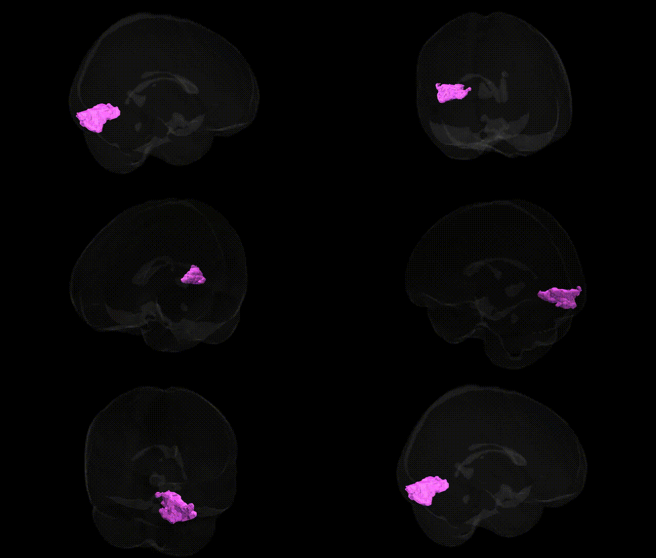
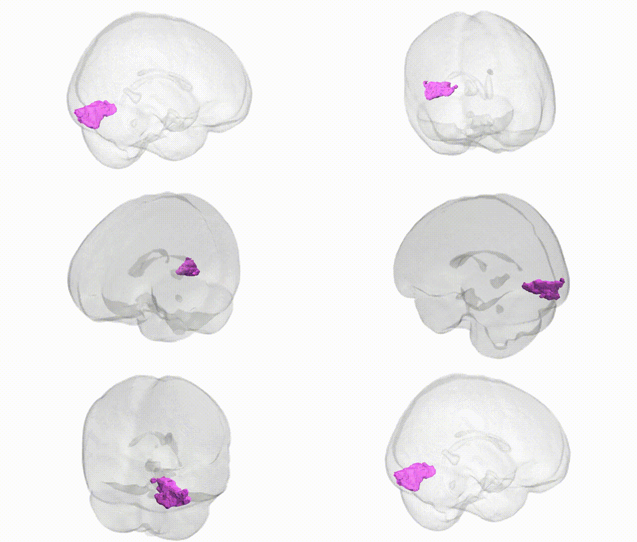
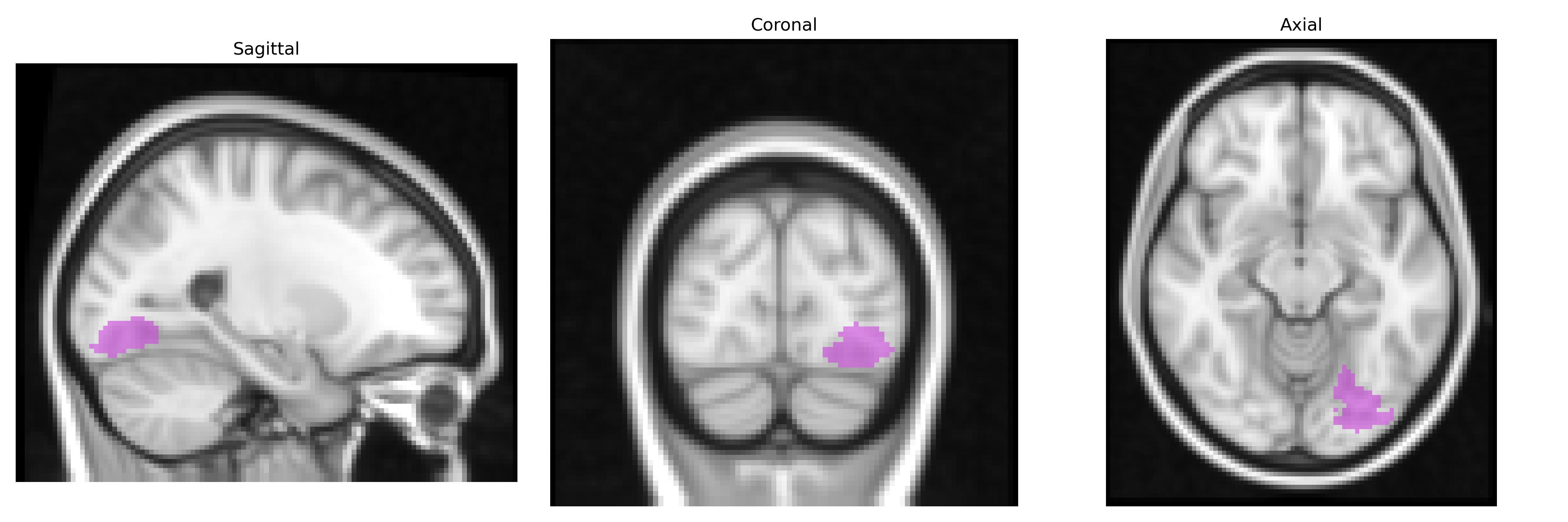
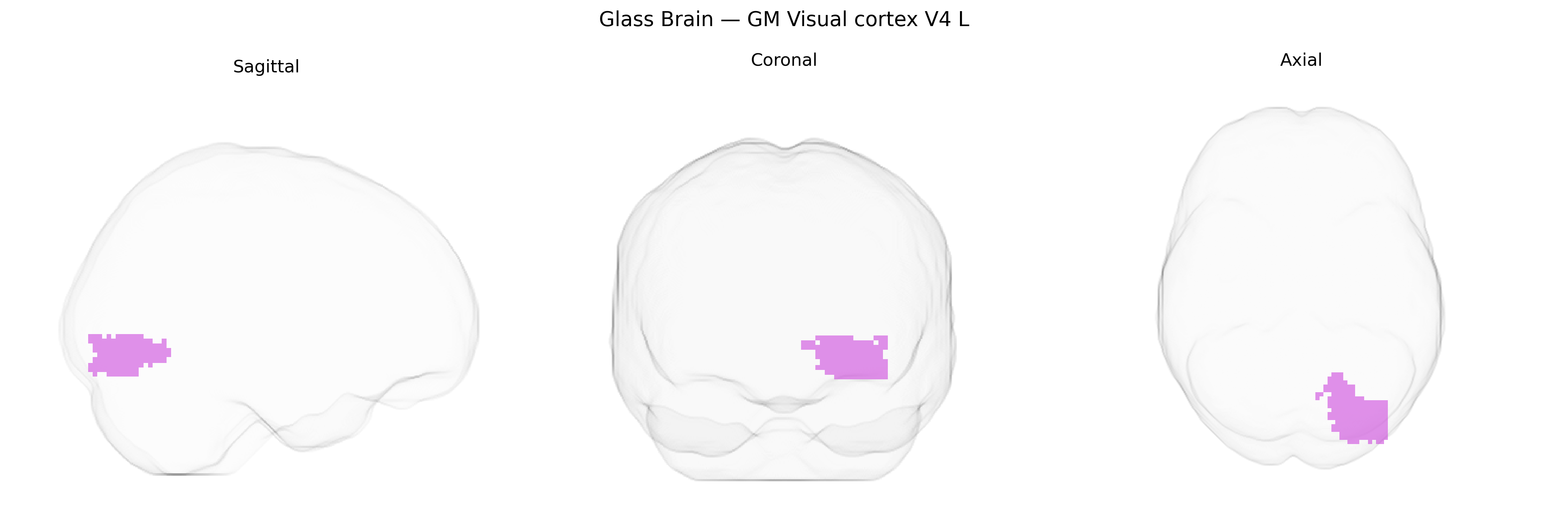
The bilateral GM Visual cortex V4 L region in the Juelich maxprob thr25 2 mm atlas corresponds to a left-hemispheric portion of area V4 within the ventral visual pathway, located on the ventrolateral surface of the occipital lobe extending into the occipitotemporal region. This area is characterized by its role in intermediate-to-higher order visual processing, particularly in the analysis of color, form, and complex shape features, and is implicated in the integration of visual inputs necessary for object recognition. Cytoarchitectonically, V4 exhibits distinct laminar organization compared with earlier visual areas (e.g., V1–V3), and functionally it shows strong connectivity with both earlier retinotopic visual areas and downstream ventral stream regions such as inferior temporal cortex. There is no direct link for “Visual cortex V4 L” as a separate entry, but it is part of the broader Visual area V4.
The bilateral GM visual cortex V4 (L) from the Juelich maxprob thr25 2 mm atlas corresponds to a mid-level extrastriate region implicated in color and form processing, and genetic associations involving this area typically arise from large-scale imaging genetics and GWAS of cortical structure and functional activation rather than region-specific studies. GWAS of occipital and visual cortical thickness, surface area, and folding (e.g., ENIGMA and UK Biobank cohorts) have repeatedly implicated loci near genes involved in neurodevelopment and synaptic function (such as variants in or near KIAA0586, CENPW, PAX6, and others regulating cortical arealization and visual system development), though these findings generally refer to broader occipital or visual association territories that include V4 rather than uniquely isolating this parcel. Polygenic influences on V4-related activation patterns have been inferred in GWAS of visual perceptual traits, including color perception and face/object recognition, where associated variants often map to genes involved in glutamatergic signaling, myelination, and neuronal migration, but precise parcel-level mapping is not yet standard. In clinical genetic studies, alterations of V4 structure and function have been reported in conditions such as developmental prosopagnosia, autism spectrum disorder, and schizophrenia, with risk loci and rare variants in genes like NRXN1, GRIN2A/B, and other synaptic genes contributing to atypical higher-order visual processing; however, these associations typically involve distributed occipito-temporal networks rather than demonstrating a unique, causally specific relationship with V4 in the Juelich atlas. Overall, genetic influences on the bilateral GM visual cortex V4 (L) are supported by broad GWAS of occipital/visual cortical anatomy and function, but current evidence links this region to polygenic neurodevelopmental and synaptic pathways shared with neighboring visual areas rather than to discrete, V4-exclusive genetic risk factors for particular disorders or traits.
Overview generated by GPT-4o (2026).
Region ID: 87
Hemisphere: bilateral
Atlas: Juelich maxprob thr25 2mm
Full Quality Version: Download MP4
Full Quality Version: Download MP4
Wali Sidiqyar*, Gaurav Rudravaram*, Elyssa M. McMaster, Trent M. Schwartz, Adam M. Saunders, Kurt G. Schilling, Bennett A. Landman "Introducing SPINS: A Shared Public Visualization Library of Neuroanatomical Structures." Medical Imaging with Deep Learning- short paper
This resource is licensed under CC0 1.0 Universal (Public Domain).