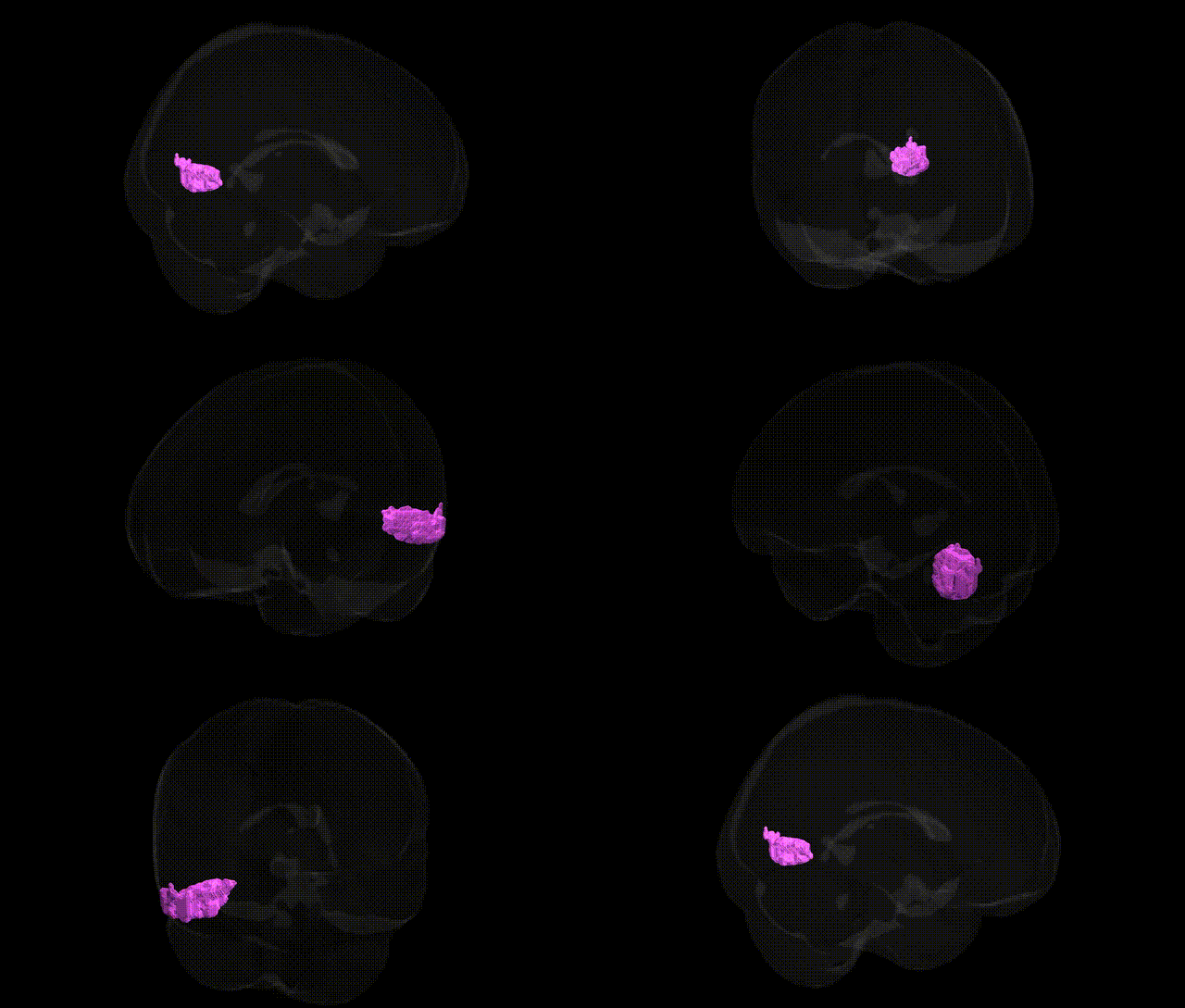
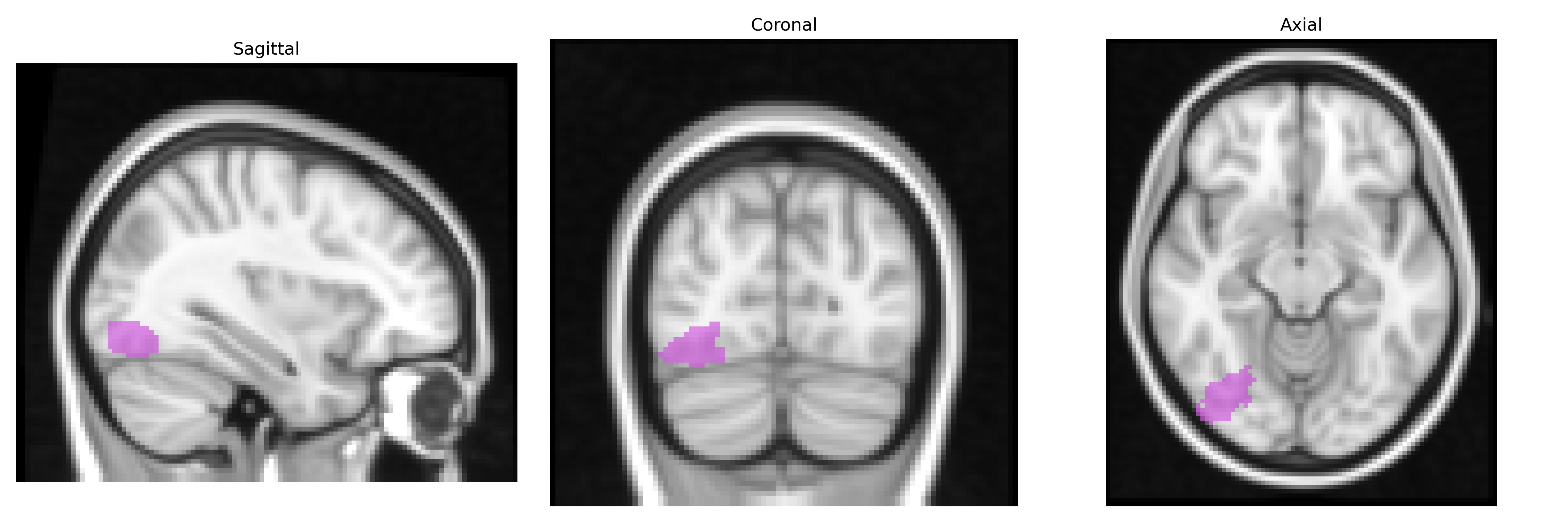
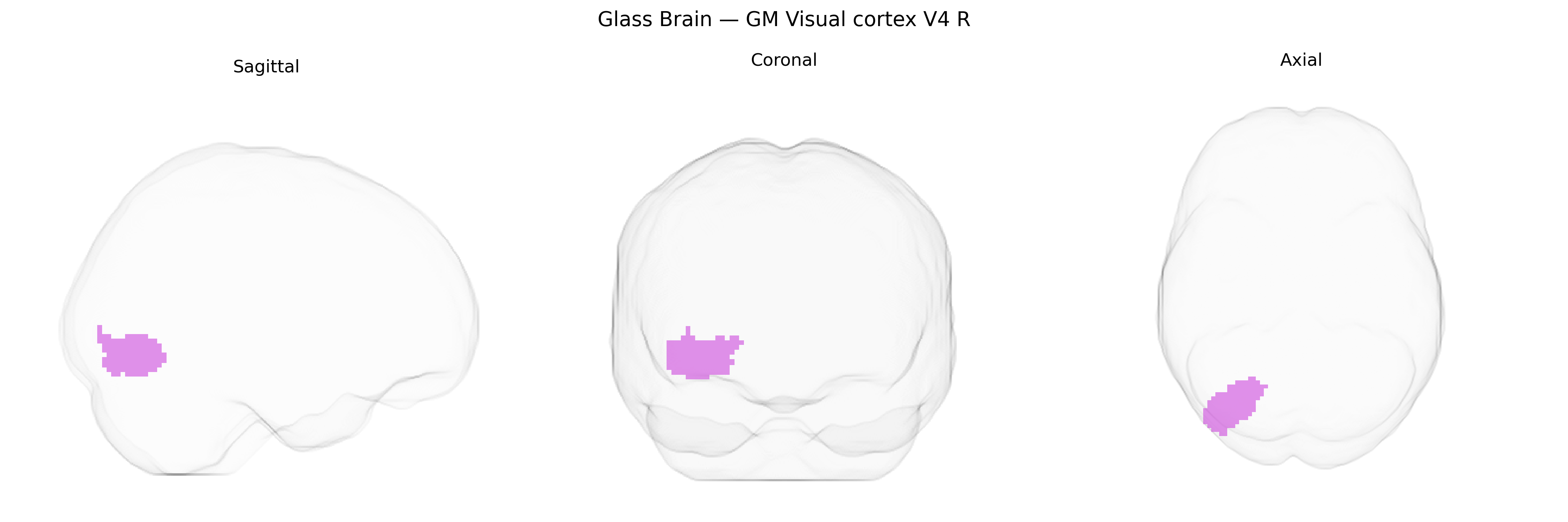
The bilateral GM Visual cortex V4 R region in the Juelich maxprob thr25 2mm atlas corresponds to right-hemisphere gray matter in human visual area V4, a mid-level extrastriate visual region located on the ventral surface of the occipital lobe, extending into the occipitotemporal cortex. V4 is part of the ventral visual stream and is critically involved in processing color, form, and complex object features, integrating inputs from earlier visual areas (such as V1 and V2) and contributing to perception of surface properties and shape-based recognition. Neurons in V4 exhibit receptive fields selective for color, curvature, and orientation, and the region shows strong modulation by attention and figure–ground segregation. In humans, V4 is commonly identified with functional imaging and retinotopic mapping; cytoarchitectonic maps such as the Juelich atlas refine its localization relative to surrounding visual areas like V3 and the lateral occipital complex. There is no direct Wikipedia article for “human V4” as defined in the Juelich atlas; a related structure and functional context is described under Visual area V4.
Genetic associations specifically targeting the bilateral GM Visual cortex V4 R region in the Juelich maxprob thr25 2mm Atlas are sparse, as most imaging genetics studies focus on broader occipital or visual cortical areas rather than cytoarchitectonic subregions like V4. However, variants in genes involved in synaptic plasticity, neurodevelopment, and visual processing—such as BDNF, GRIN2B, and genes from the glutamatergic and GABAergic pathways—have been linked to structural and functional variation across the visual cortex in general, including regions overlapping V4. GWAS of cortical thickness and surface area (e.g., ENIGMA and UK Biobank studies) have identified multiple loci (including variants near genes such as TBR1, PAX6, and FOXP1) that influence occipital cortex morphology, which likely encompass V4 given the spatial resolution of these analyses. Additionally, genetic studies of disorders with known visual-processing abnormalities, such as autism spectrum disorder, schizophrenia, and dyslexia, have implicated polygenic risk scores that correlate with altered visual cortical activation patterns and connectivity, sometimes extending into lateral occipital and V4-like territories. GWAS of traits related to visual perception (e.g., color vision, face recognition, and migraine with aura) have identified loci in pathways for retinal and cortical visual processing (including genes like OPN1LW/OPN1MW for color vision and CACNA1A for migraine) that are functionally connected to extrastriate cortex, though these studies rarely isolate V4 as a distinct anatomical target. Overall, genetic findings point to a polygenic influence on occipital/visual cortex structure and function, with indirect but plausible relevance to the V4 region defined in the Juelich atlas, rather than direct, region-specific GWAS hits.
Overview generated by GPT-4o (2026).
Region ID: 88
Hemisphere: bilateral
Atlas: Juelich maxprob thr25 2mm
Full Quality Version: Download MP4
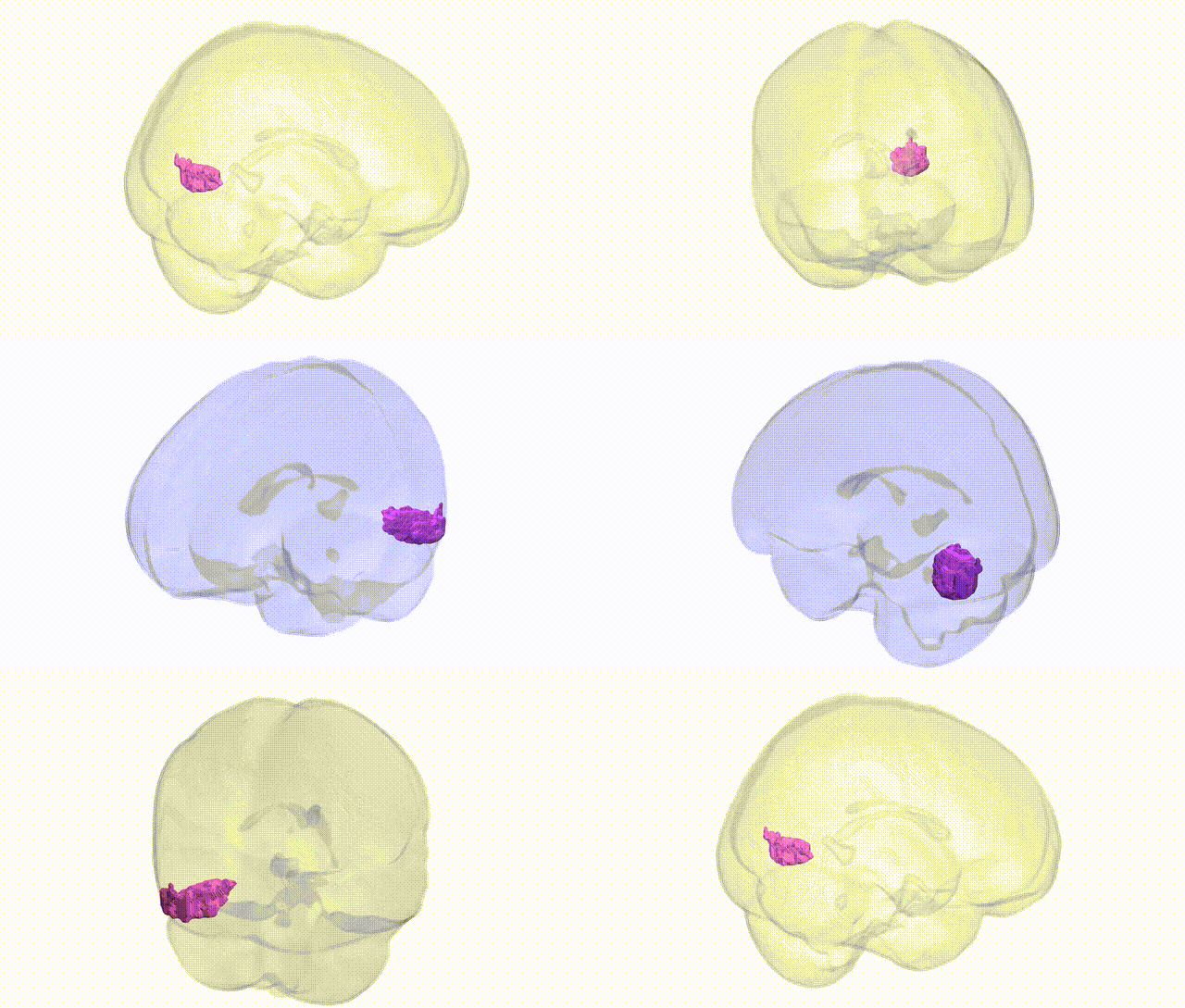
Full Quality Version: Download MP4
Wali Sidiqyar*, Gaurav Rudravaram*, Elyssa M. McMaster, Trent M. Schwartz, Adam M. Saunders, Kurt G. Schilling, Bennett A. Landman "Introducing SPINS: A Shared Public Visualization Library of Neuroanatomical Structures." Medical Imaging with Deep Learning- short paper
This resource is licensed under CC0 1.0 Universal (Public Domain).