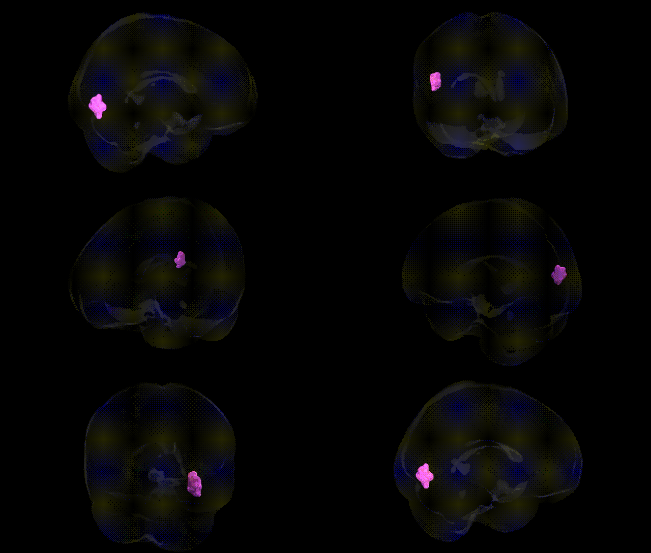
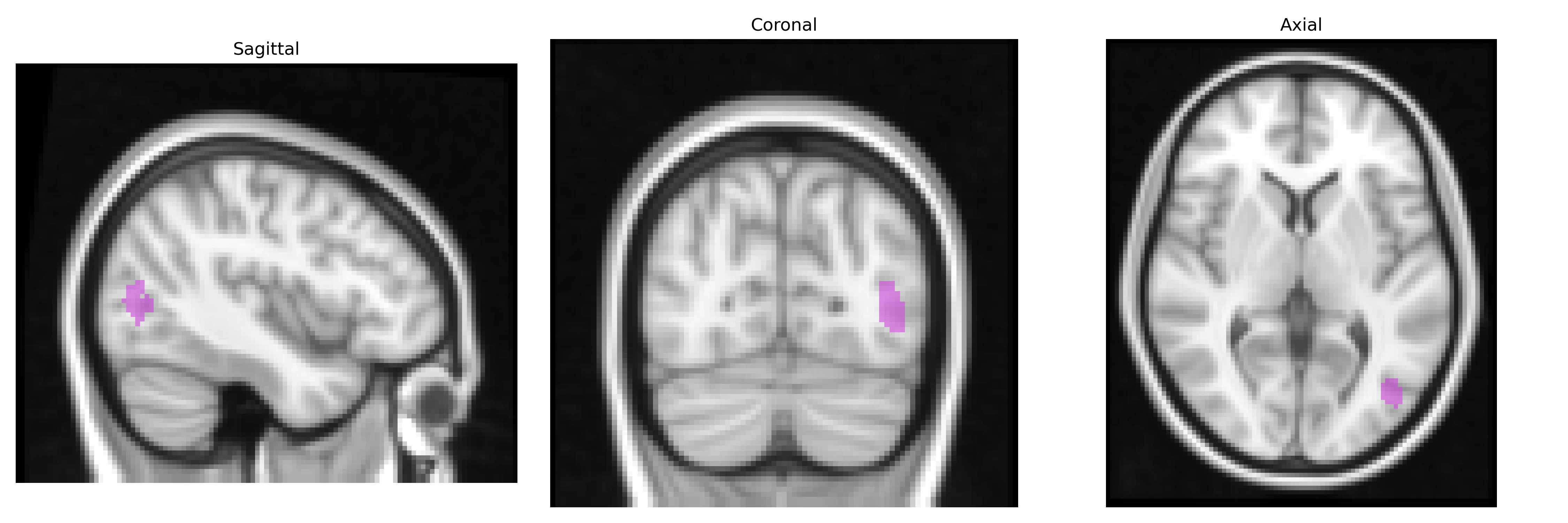
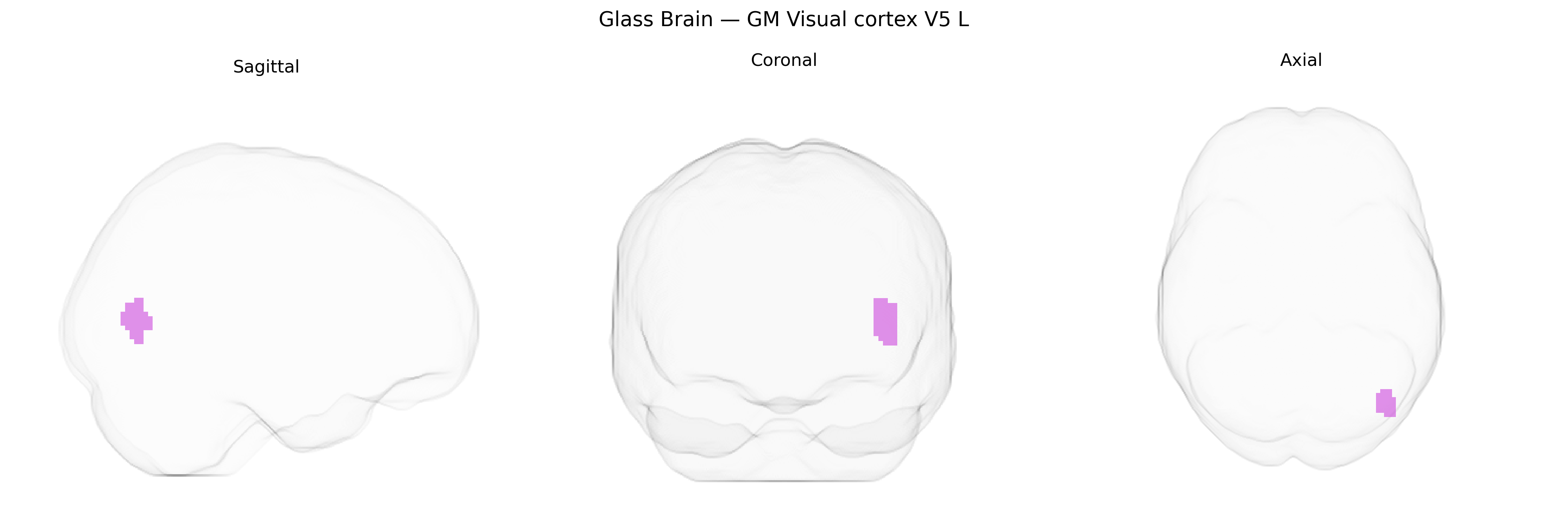
The bilateral GM Visual cortex V5 L region from the Juelich maxprob thr25 2mm atlas corresponds to the left hemisphere portion of human visual area V5/MT, a motion-sensitive extrastriate visual region located in the lateral occipitotemporal cortex. This area is critically involved in the analysis of visual motion, including detection of direction, speed, and coherence of moving stimuli, and contributes to motion perception, smooth pursuit eye movements, and visuomotor integration. Neurons in V5/MT exhibit strong direction selectivity and integrate information from earlier visual areas such as V1 and V2, while also interacting with parietal and frontal regions involved in attention and action planning. As part of the dorsal visual stream, V5/MT supports the transformation of dynamic visual information into spatial and movement-related representations important for navigation and object tracking. There is no direct Wikipedia article for “Visual cortex V5 L”; a related article describing this functional complex is Visual area MT.
The bilateral GM Visual cortex V5 (L), corresponding to human MT/V5 in the Juelich maxprob atlas, is a motion-selective extrastriate visual area for which direct, region-specific genetic associations are still limited, but broader imaging-genetics and GWAS work implicates several pathways influencing its structure and function. Large-scale brain MRI GWAS (e.g., ENIGMA, UK Biobank) have identified common variants in genes involved in synaptic development, cortical patterning, and myelination—such as BDNF, NTRK2, CNTNAP2, and genes in the Wnt and MAPK pathways—as contributors to inter-individual variation in occipito-temporal cortical thickness and surface area, including regions overlapping MT/V5. Polygenic risk for schizophrenia, bipolar disorder, autism spectrum disorder, and major depression has been associated with altered activation and connectivity in motion-sensitive visual cortex during visual processing and eye-movement tasks, suggesting that shared neurodevelopmental risk variants affect this region as part of larger dorsal visual and frontoparietal networks. GWAS of visual perception and reading-related traits (including dyslexia) have implicated genes such as DCDC2 and KIAA0319, with downstream effects on magnocellular/dorsal stream circuits that encompass MT/V5, and genetic factors influencing white-matter integrity (e.g., CNTN4, LINGO1) have been linked to fractional anisotropy in tracts connecting posterior occipital regions to MT/V5. Additionally, variants affecting glutamatergic and GABAergic signaling (such as in GRIN2B and GABRA genes) have been associated with motion perception thresholds and visual cortical excitability, indirectly implicating MT/V5 circuitry, while migraine and visual aura GWAS (e.g., involving TRPM8, PHACTR1) point to vascular and excitability-related mechanisms that influence susceptibility to visual motion disturbances engaging this area.
Overview generated by GPT-4o (2026).
Region ID: 89
Hemisphere: bilateral
Atlas: Juelich maxprob thr25 2mm
Full Quality Version: Download MP4
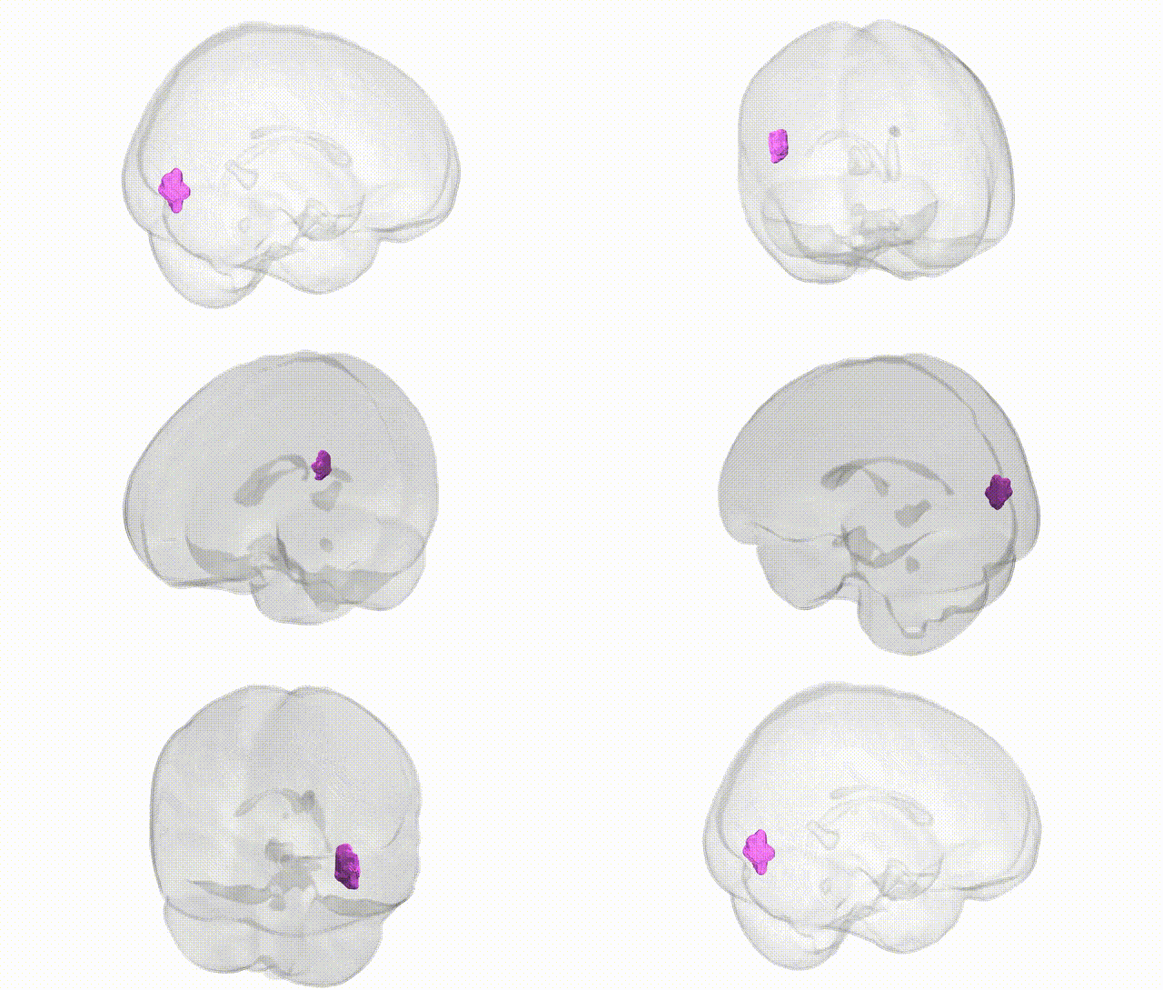
Full Quality Version: Download MP4
Wali Sidiqyar*, Gaurav Rudravaram*, Elyssa M. McMaster, Trent M. Schwartz, Adam M. Saunders, Kurt G. Schilling, Bennett A. Landman "Introducing SPINS: A Shared Public Visualization Library of Neuroanatomical Structures." Medical Imaging with Deep Learning- short paper
This resource is licensed under CC0 1.0 Universal (Public Domain).