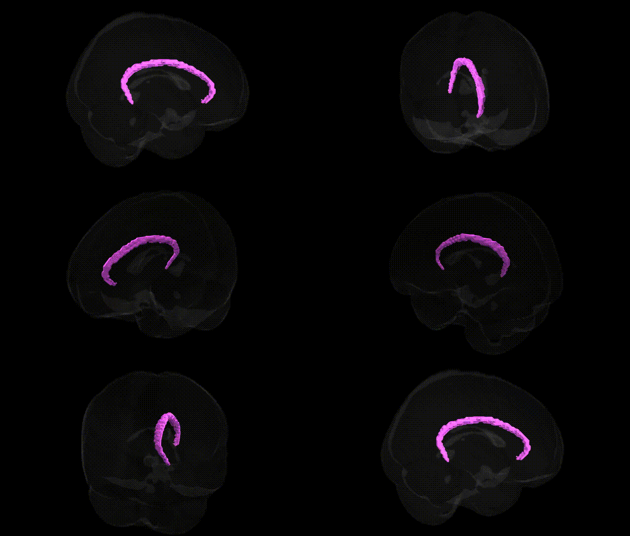
The bilateral WM Cingulum L from the Juelich maxprob thr25 2mm Atlas refers to the left-side white matter portion of the cingulum bundle, a major association fiber tract that runs within the cingulate gyrus and connects medial frontal, parietal, and temporal lobe structures, including components of the limbic system such as the anterior cingulate cortex, posterior cingulate cortex, and hippocampal formation. This tract supports integration of cognitive, emotional, and mnemonic processes by facilitating communication among regions involved in attention, executive control, emotion regulation, and episodic memory. Its integrity is crucial for efficient network-level functioning of the default mode and limbic circuits, and alterations in the cingulum bundle have been implicated in various neuropsychiatric and neurodegenerative conditions. There is no direct link for “WM Cingulum L”; a related structure is the Cingulum (brain).
The bilateral white-matter cingulum (particularly the cingulum bundle underlying the left cingulate cortex as defined in the Juelich maxprob thr25 2 mm atlas) shows heritable variation in microstructure and volume, with SNP-based heritability estimates from large diffusion MRI GWAS (e.g., UK Biobank) indicating moderate genetic influence on fractional anisotropy and mean diffusivity, and polygenic overlap with general cognitive ability, educational attainment, and psychiatric phenotypes. Multiple studies have identified associations between cingulum white-matter integrity and common variants in genes implicated in neurodevelopment, myelination, and synaptic function, including BDNF (e.g., Val66Met), NRG1/ErbB signaling components, DISC1-related pathways, and genes affecting oligodendrocyte biology, although individual locus effects are generally small and often not specific to this tract alone. GWAS of major depressive disorder, schizophrenia, bipolar disorder, anxiety, and autism spectrum disorder converge on networks involving the cingulum and broader limbic white-matter, with polygenic risk scores for these conditions correlating with altered cingulum FA or connectivity, particularly in anterior and dorsal subdivisions. In Alzheimer’s disease and other dementias, risk alleles in APOE and additional loci identified by large AD GWAS show downstream associations with cingulum degeneration and disrupted default-mode connectivity, while imaging-genetics studies in normal aging link cingulum changes to variants in genes regulating amyloid processing, tau, and vascular risk. Collectively, genetic findings suggest that the bilateral cingulum is a key intermediate phenotype integrating polygenic risk for mood and psychotic disorders, cognitive performance, and neurodegeneration, mediated by genes that influence axonal organization, myelination, and plasticity in limbic–frontal circuits.
Overview generated by GPT-4o (2026).
Region ID: 97
Hemisphere: bilateral
Atlas: Juelich maxprob thr25 2mm
Full Quality Version: Download MP4
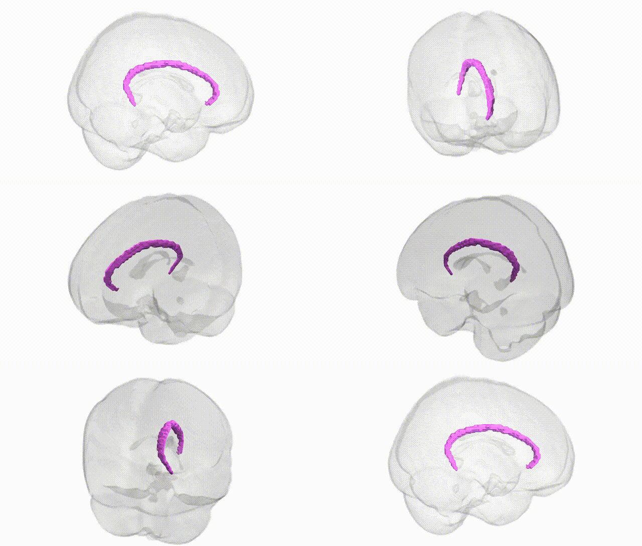
Full Quality Version: Download MP4
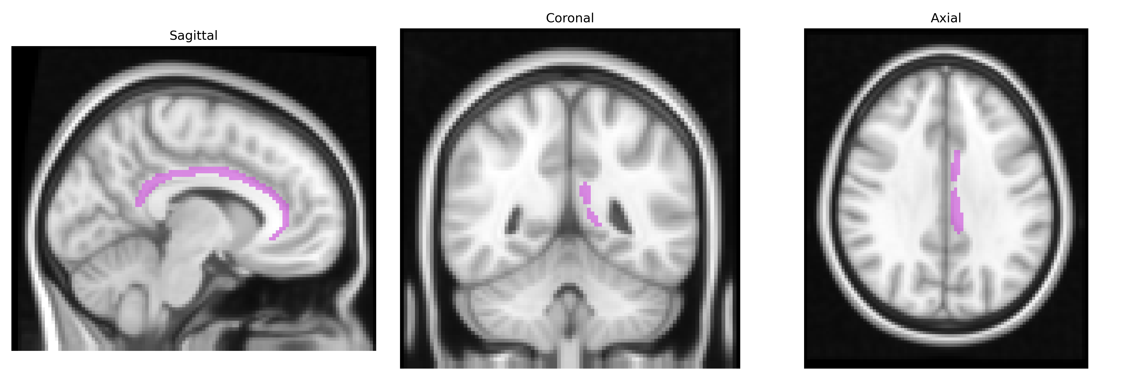
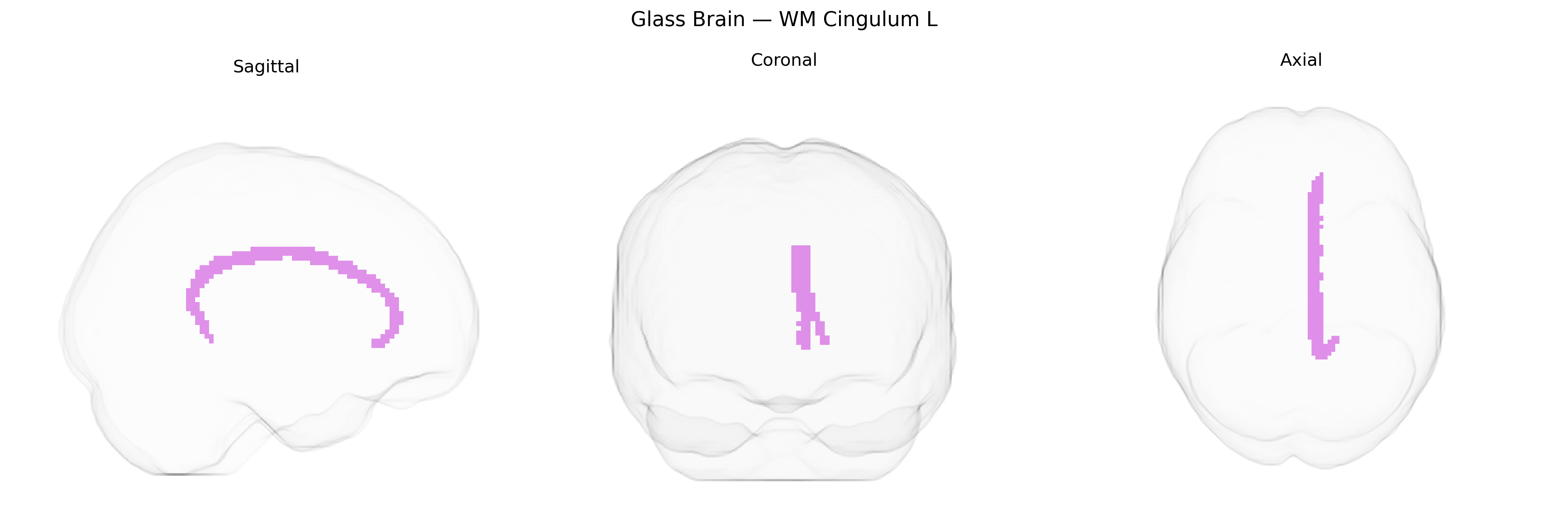
Wali Sidiqyar*, Gaurav Rudravaram*, Elyssa M. McMaster, Trent M. Schwartz, Adam M. Saunders, Kurt G. Schilling, Bennett A. Landman "Introducing SPINS: A Shared Public Visualization Library of Neuroanatomical Structures." Medical Imaging with Deep Learning- short paper
This resource is licensed under CC0 1.0 Universal (Public Domain).