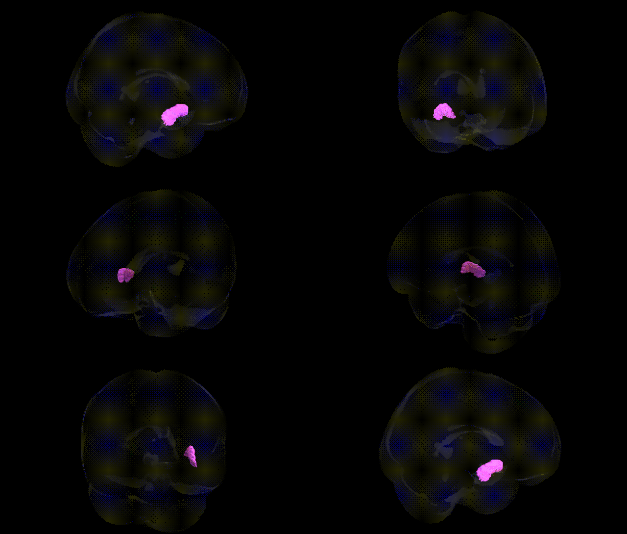
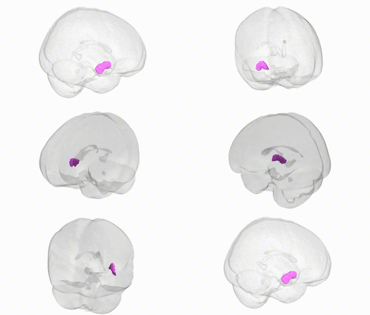
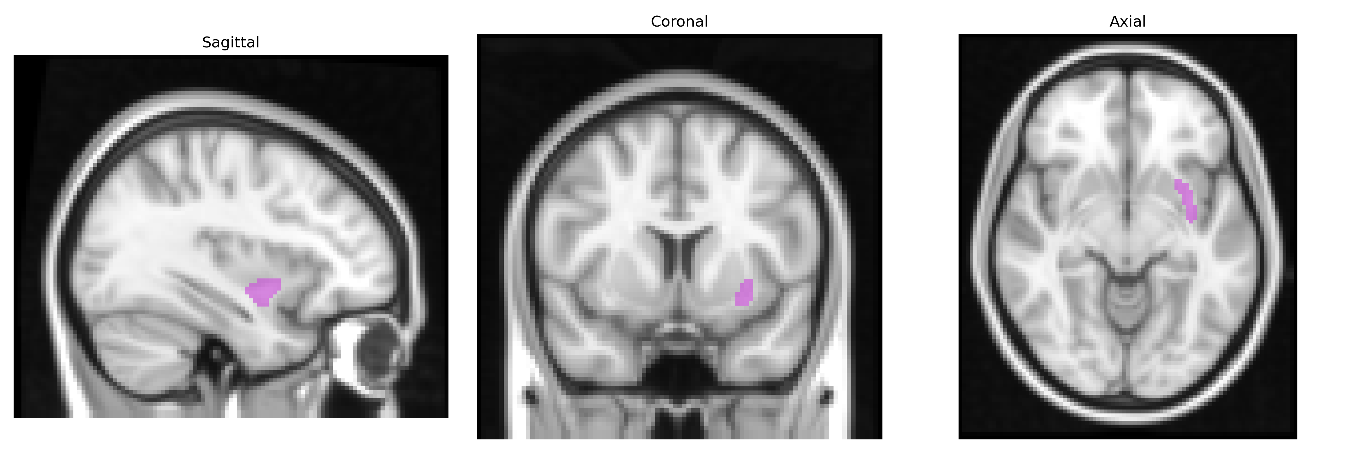
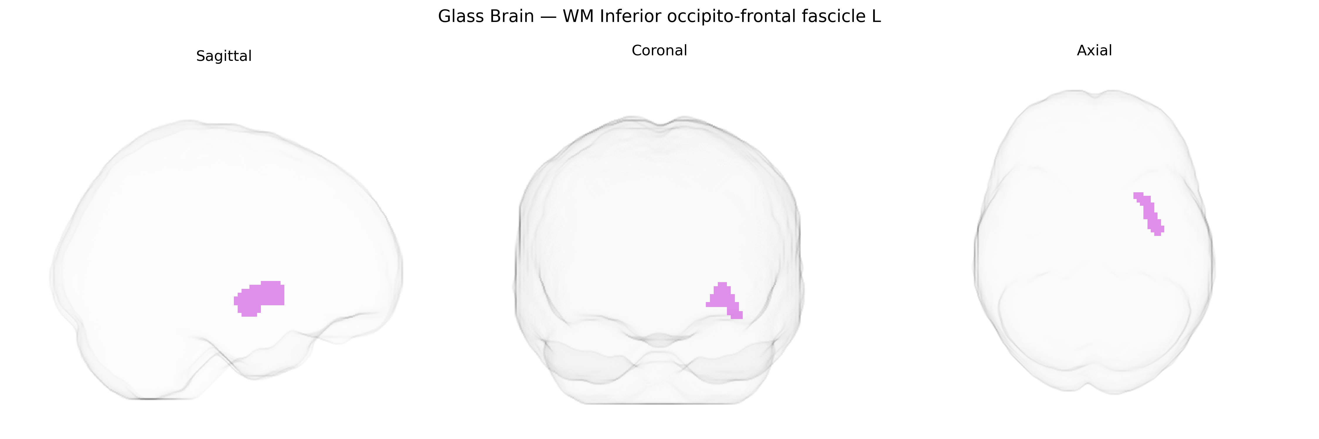
The bilateral WM Inferior occipito-frontal fascicle L from the Juelich maxprob thr25 2mm Atlas corresponds to the left inferior fronto-occipital fasciculus (IFOF), a major associative white matter tract that links occipital and posterior temporal regions with the frontal lobe. This fascicle courses through the deep white matter of the temporal and parietal lobes, typically passing medial to the insula and through the external and extreme capsules, supporting long-range integration between visual, semantic, and higher-order cognitive networks. Functionally, the IFOF has been implicated in language processing, semantic comprehension, visual recognition, and aspects of attention, and is often studied via diffusion MRI tractography and lesion-symptom mapping in patients with stroke, tumors, or neurodegenerative disease. There is no direct Wikipedia article for this exact atlas label; a closely related structure is the Inferior fronto-occipital fasciculus.
The bilateral inferior fronto‑occipital fasciculus (IFOF) white matter tract, as defined in the Juelich maxprob thr25 2 mm atlas, has been implicated in several genetic and imaging‑genetics studies, although direct, tract‑specific GWAS findings are limited and often embedded within broader measures of white matter integrity. Large neuroimaging GWAS consortia such as ENIGMA and UK Biobank have identified common variants associated with diffusion tensor imaging (DTI) metrics (e.g., fractional anisotropy, mean diffusivity) in association fibers that include the IFOF, with prominent signals near genes involved in axon guidance, myelination, and neurodevelopment (such as CNTNAP2, NRG1, and various cell adhesion and cytoskeletal genes). Polygenic risk scores for schizophrenia, bipolar disorder, and major depressive disorder have been linked to altered microstructure in IFOF and adjacent tracts, suggesting that shared genetic liability for these disorders may partly act through disruption of long‑range fronto‑occipital connectivity. Similarly, variants associated with autism spectrum disorder and attention‑deficit/hyperactivity disorder show convergent evidence of altered IFOF integrity, in line with the tract’s role in social cognition, language, and attention. In neurodegenerative disease, Alzheimer’s disease‑related genetic risk (including APOE ε4) has been associated with changes in association white matter, potentially involving the IFOF, although effects are often distributed and not specific to this tract. Overall, current evidence points to the IFOF as a genetically modulated conduit for higher‑order cognitive and psychiatric phenotypes, but most findings derive from global or multi‑tract white matter GWAS rather than region‑exclusive analyses, and the Juelich atlas label serves primarily as an anatomical reference within these broader imaging‑genetics frameworks.
Overview generated by GPT-4o (2026).
Region ID: 102
Hemisphere: bilateral
Atlas: Juelich maxprob thr25 2mm
Full Quality Version: Download MP4
Full Quality Version: Download MP4
Wali Sidiqyar*, Gaurav Rudravaram*, Elyssa M. McMaster, Trent M. Schwartz, Adam M. Saunders, Kurt G. Schilling, Bennett A. Landman "Introducing SPINS: A Shared Public Visualization Library of Neuroanatomical Structures." Medical Imaging with Deep Learning- short paper
This resource is licensed under CC0 1.0 Universal (Public Domain).