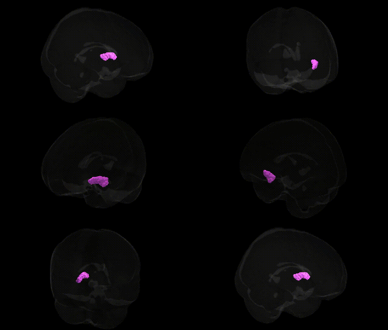
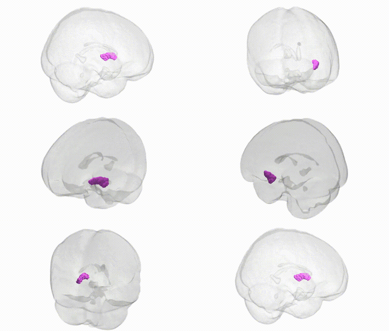
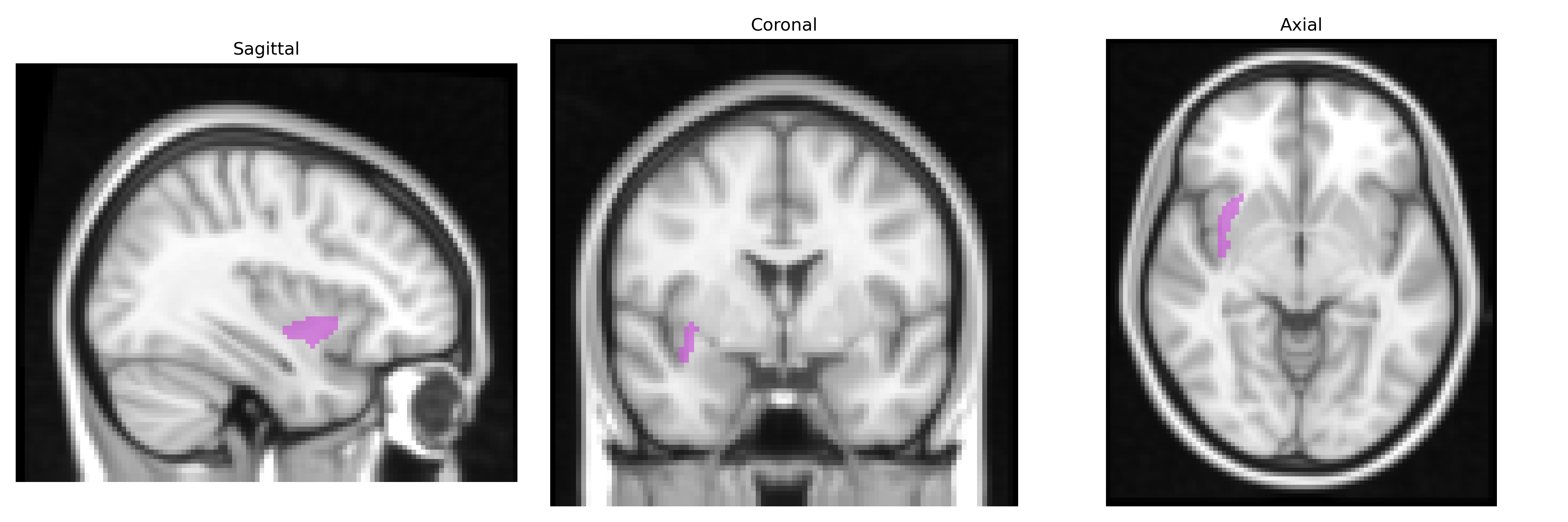
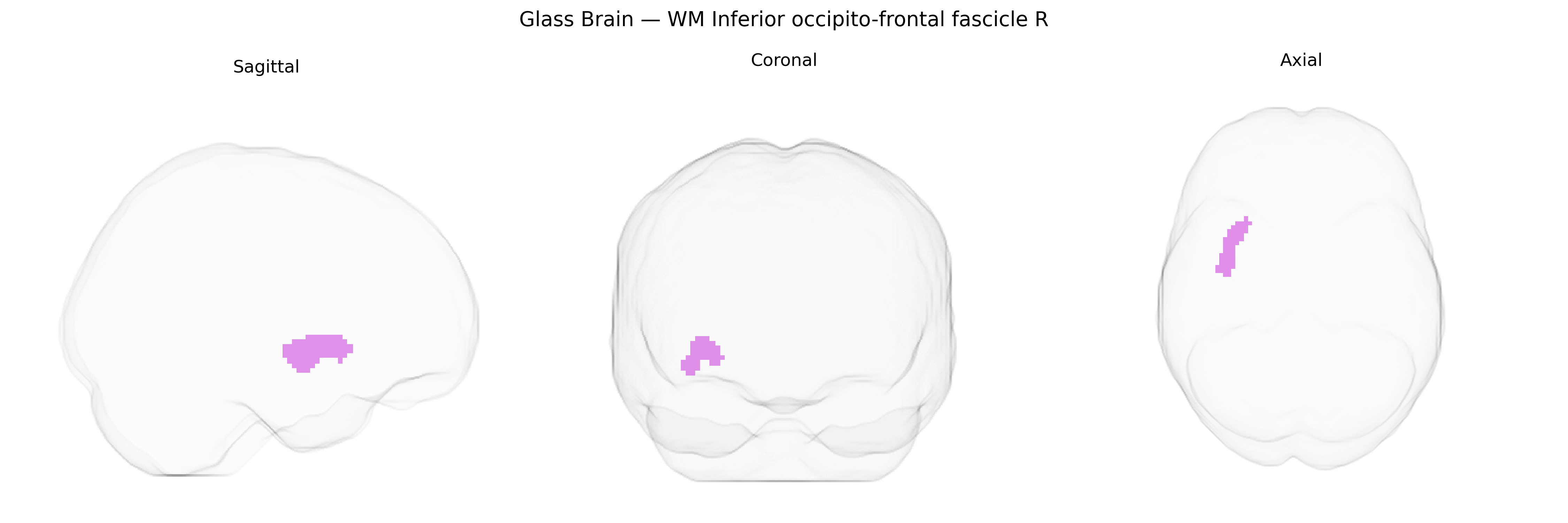
The bilateral WM Inferior occipito-frontal fascicle R in the Juelich maxprob thr25 2mm atlas corresponds to the inferior fronto-occipital fasciculus (IFOF), a major associative white matter tract connecting occipital and posterior temporal regions with frontal cortices via the ventral external capsule and deep white matter. It is implicated in the integration of visual information with higher-order cognitive functions, including semantic processing, language, attention, and aspects of executive control, by mediating long-range communication between visual association areas, the temporal lobe, and prefrontal regions. Structurally, the IFOF courses medially to the insula and basal ganglia, with right-hemisphere fibers contributing to visuospatial and attentional networks as part of a broader ventral stream connectivity system. There is no direct Wikipedia article for this specific atlas label; a related structure is the Inferior fronto-occipital fasciculus.
The right inferior fronto-occipital fasciculus (IFOF) is a major association white-matter tract implicated in language, visual processing, and higher-order cognition, and genetic studies of white-matter microstructure and connectivity have identified multiple loci influencing its integrity. Large diffusion MRI GWAS (e.g., ENIGMA, UK Biobank) report associations between IFOF-related fractional anisotropy and genes involved in axon guidance, myelination, and cytoskeletal dynamics, including variants near or within CNTN4, NTRK3, ROBO1/2, and genes regulating oligodendrocyte function; many of these loci show pleiotropy with global white-matter metrics rather than IFOF-specific effects. Polygenic risk for schizophrenia, bipolar disorder, and major depressive disorder has been associated with reduced integrity of IFOF segments, and genes such as ZNF804A, NRG1, and DISC1 have been linked indirectly through broader fronto-temporal white-matter alterations that encompass the IFOF. Autism spectrum disorder and ADHD risk variants in synaptic and neurodevelopmental genes (e.g., CNTNAP2, MET, and DCC-related pathways) have also been tied to altered long-range association tracts including the IFOF. In Alzheimer’s disease and other dementias, APOE ε4 status and loci identified by neurodegeneration GWAS correlate with degeneration of posterior-anterior association fibers, with right IFOF involvement in networks subserving visuospatial and attentional functions. Overall, genetic effects on the bilateral inferior fronto-occipital fasciculus are distributed across many loci with modest effect sizes and tend to reflect shared neurodevelopmental and myelination biology rather than region-specific genes, with links to psychiatric disorders, neurodevelopmental conditions, and cognitive and imaging-derived brain-structure traits.
Overview generated by GPT-4o (2026).
Region ID: 101
Hemisphere: bilateral
Atlas: Juelich maxprob thr25 2mm
Full Quality Version: Download MP4
Full Quality Version: Download MP4
Wali Sidiqyar*, Gaurav Rudravaram*, Elyssa M. McMaster, Trent M. Schwartz, Adam M. Saunders, Kurt G. Schilling, Bennett A. Landman "Introducing SPINS: A Shared Public Visualization Library of Neuroanatomical Structures." Medical Imaging with Deep Learning- short paper
This resource is licensed under CC0 1.0 Universal (Public Domain).