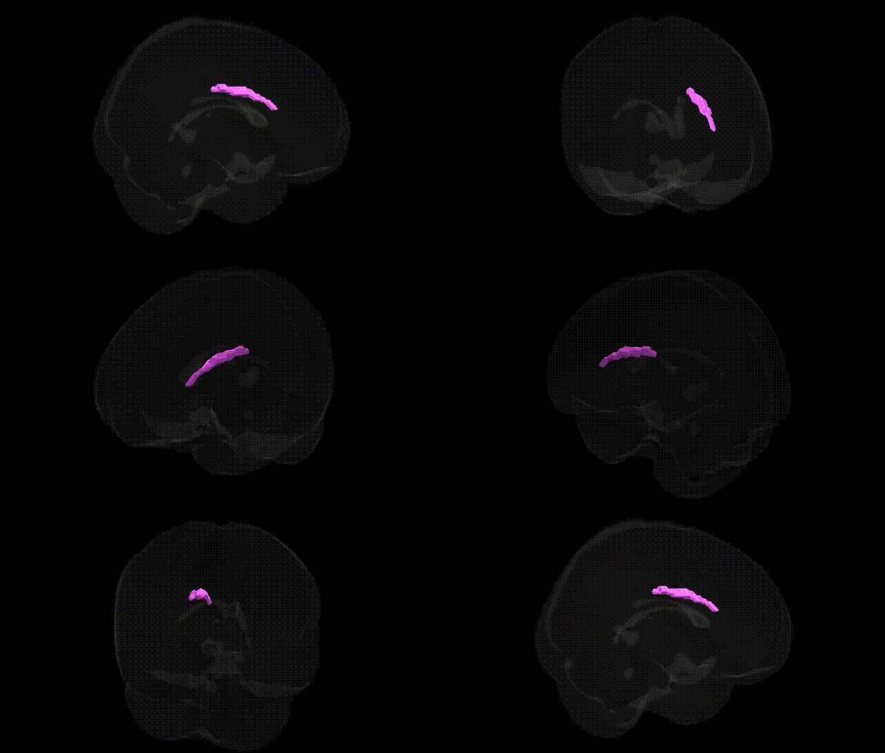
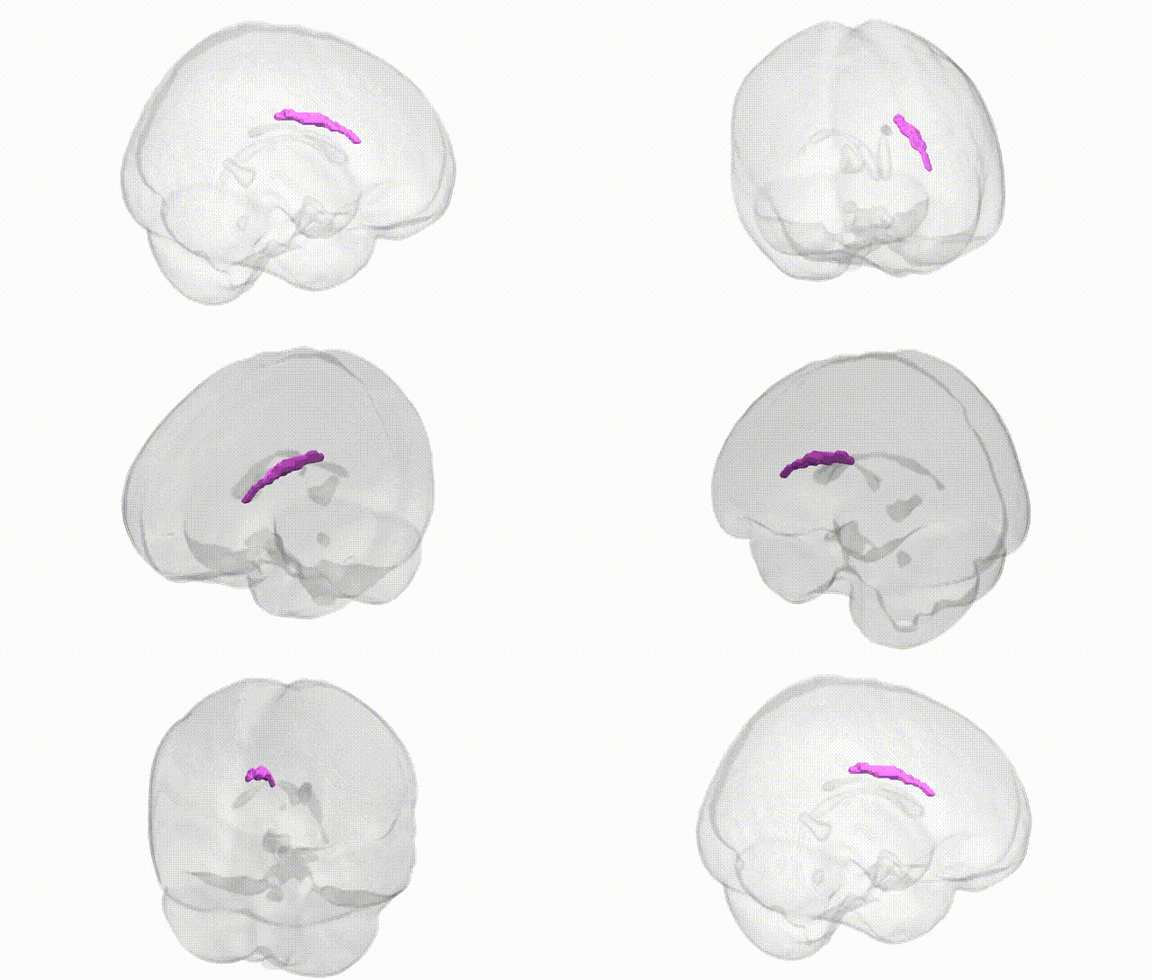
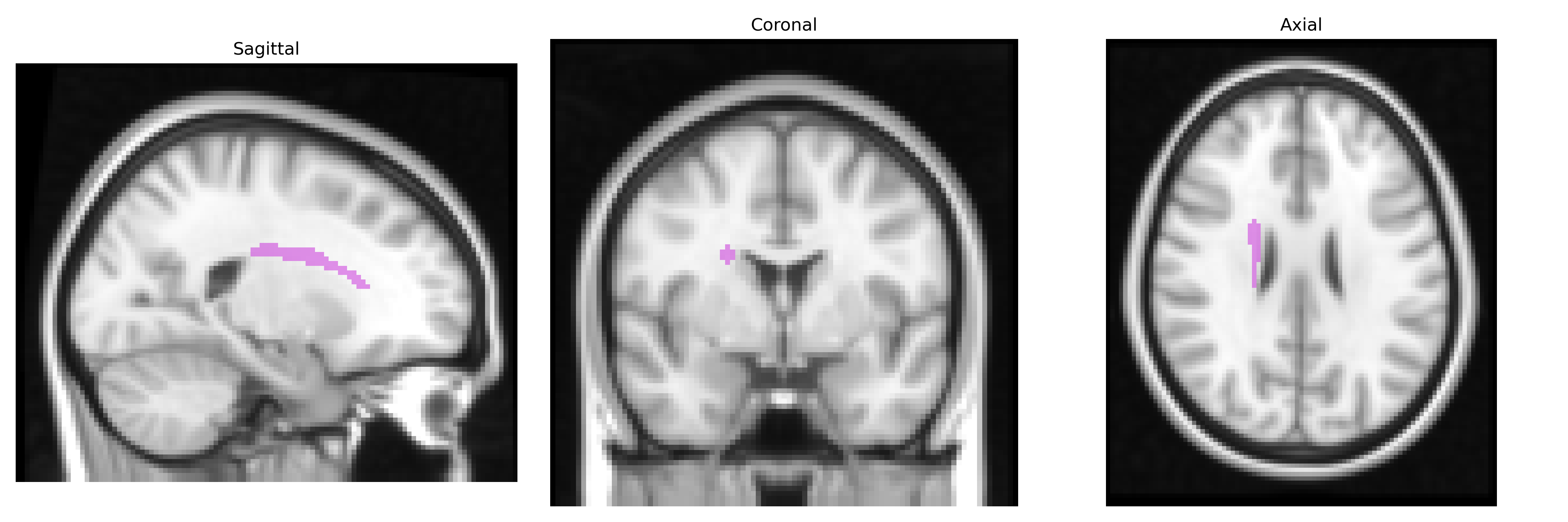
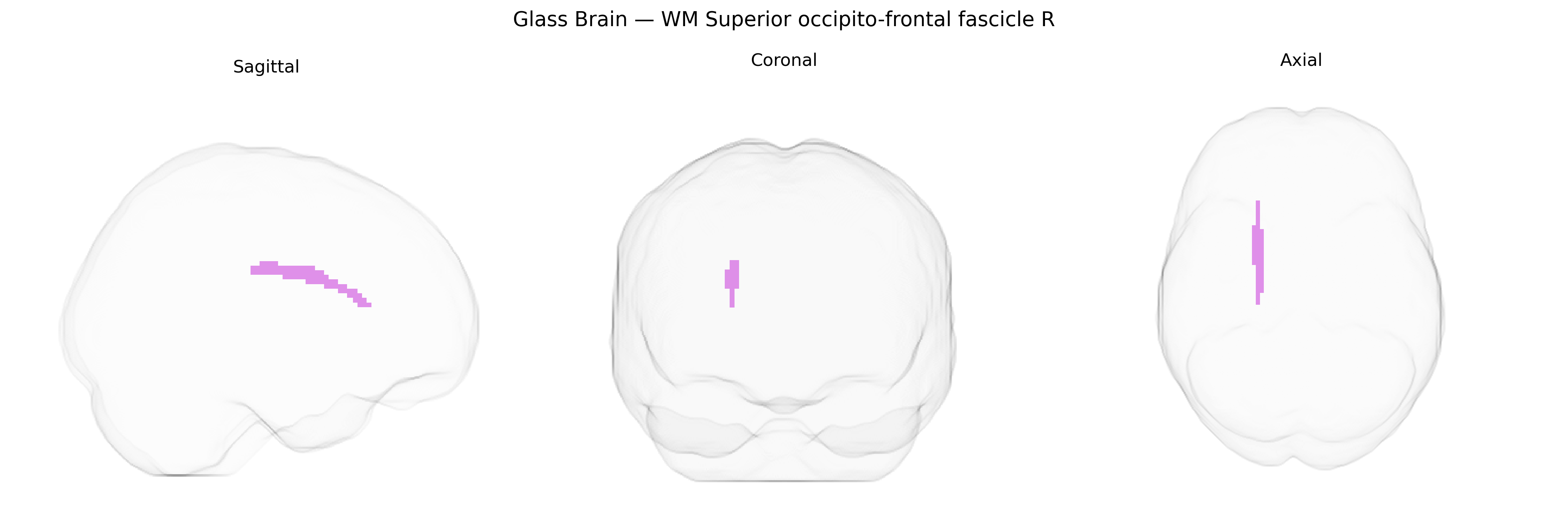
The bilateral WM Superior occipito-frontal fascicle R, as defined in the Juelich maxprob thr25 2 mm atlas, corresponds to the right superior fronto‑occipital fasciculus, a major long association white matter tract connecting dorsal and medial occipital regions with superior and medial frontal cortices via the parietal lobe and deep periventricular white matter. This fascicle supports integration of visual, spatial, and higher‑order cognitive functions by enabling rapid bidirectional communication between posterior visual areas and frontal executive and attentional networks. Anatomically, it courses lateral to the lateral ventricle and interdigitates with neighboring association fibers, including components of the superior longitudinal system. There is no direct Wikipedia entry for this exact white matter tract label; a related structure and context can be found under Frontal lobe.
The bilateral WM Superior occipito-frontal fascicle (right) in the Juelich maxprob thr25 2mm atlas corresponds closely to components of the superior longitudinal fasciculus and fronto‑occipital association tracts, for which genetic influences have been studied using diffusion MRI in large cohorts. Twin and family studies show high heritability of fractional anisotropy and other microstructural measures in these tracts, and GWAS have implicated variants near genes involved in axon guidance, myelination, and oligodendrocyte function (such as CNTN4/5, NRXN1, MAG, and MOBP) in white matter integrity in fronto‑parietal and fronto‑occipital pathways. Polygenic risk for schizophrenia, bipolar disorder, and major depressive disorder has been associated with altered diffusion metrics in superior longitudinal and fronto‑occipital fasciculi, and risk variants in genes like CACNA1C and ZNF804A have been linked to microstructural abnormalities in connected fronto‑parietal networks. Neurodevelopmental disorder risk (including autism and ADHD) shows convergent associations with these tracts through both common variants and rare copy‑number variants affecting synaptic and axon guidance genes. In addition, GWAS of cognitive ability, educational attainment, and processing speed have repeatedly identified loci whose effects are partly mediated by white matter structure in long association fibers, including the superior occipito‑frontal connections represented in the Juelich atlas, underscoring a genetically influenced role of this region in higher cognition and vulnerability to psychiatric illness.
Overview generated by GPT-4o (2026).
Region ID: 112
Hemisphere: bilateral
Atlas: Juelich maxprob thr25 2mm
Full Quality Version: Download MP4
Full Quality Version: Download MP4
Wali Sidiqyar*, Gaurav Rudravaram*, Elyssa M. McMaster, Trent M. Schwartz, Adam M. Saunders, Kurt G. Schilling, Bennett A. Landman "Introducing SPINS: A Shared Public Visualization Library of Neuroanatomical Structures." Medical Imaging with Deep Learning- short paper
This resource is licensed under CC0 1.0 Universal (Public Domain).