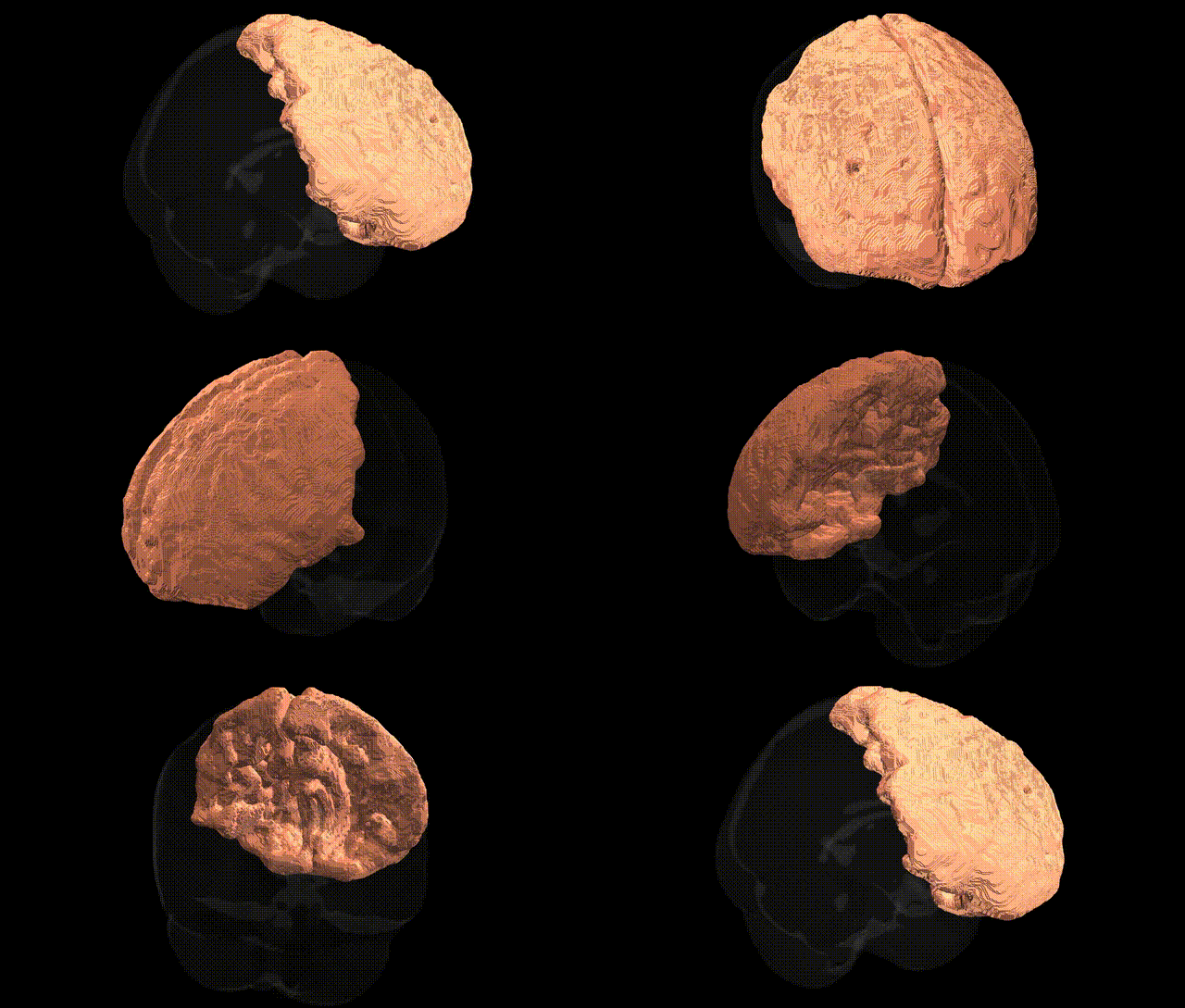
The bilateral frontal lobe, as defined in the MNI Structural maxprob thr25 1 mm atlas, comprises the anterior portion of the cerebral hemispheres, extending from the central sulcus to the frontal pole and encompassing regions involved in higher-order cognitive, motor, and behavioral functions. It contains primary and premotor cortices, prefrontal regions (including dorsolateral, ventromedial, and orbitofrontal areas), and is critically involved in executive functions such as planning, decision-making, working memory, cognitive control, and the regulation of social and emotional behavior. The frontal lobe also houses Broca’s area (typically in the dominant hemisphere) for language production and contributes to voluntary motor control via corticospinal projections originating from the precentral gyrus. Developmentally, this region is among the last to fully mature, and structurally it is organized into multiple cytoarchitectonic fields that support the integration of sensory, limbic, and motor information necessary for goal-directed behavior. Frontal lobe
The bilateral frontal lobe, as defined in the MNI Structural maxprob thr25 1mm atlas, has been implicated in numerous genetic associations identified through GWAS and imaging-genetics studies, largely via measures of cortical thickness, surface area, and volume. Common variants in genes involved in synaptic function, neurodevelopment, and cell adhesion (e.g., BDNF, GRIN2B, CNTNAP2, DISC1, CACNA1C) have been linked to structural and functional variation in frontal regions, which in turn associate with cognitive traits such as general intelligence, executive function, working memory, and educational attainment. Polygenic risk for psychiatric disorders including schizophrenia, bipolar disorder, major depressive disorder, ADHD, and autism spectrum disorder shows consistent relationships with frontal lobe morphology and connectivity, with frontal deficits frequently mediating genetic risk for impaired cognitive control and emotion regulation. Large-scale GWAS of brain imaging phenotypes (e.g., ENIGMA, UK Biobank) have identified multiple loci influencing frontal cortical thickness and volume, some overlapping with genes associated with neurodevelopmental disorders, Alzheimer’s disease, and other neurodegenerative conditions, supporting a shared genetic architecture linking frontal lobe structure to both normal variation in cognition and vulnerability to psychiatric and neurological disease.
Overview generated by GPT-4o (2026).
Region ID: 3
Hemisphere: bilateral
Atlas: MNI Structural maxprob thr25 1mm
Full Quality Version: Download MP4
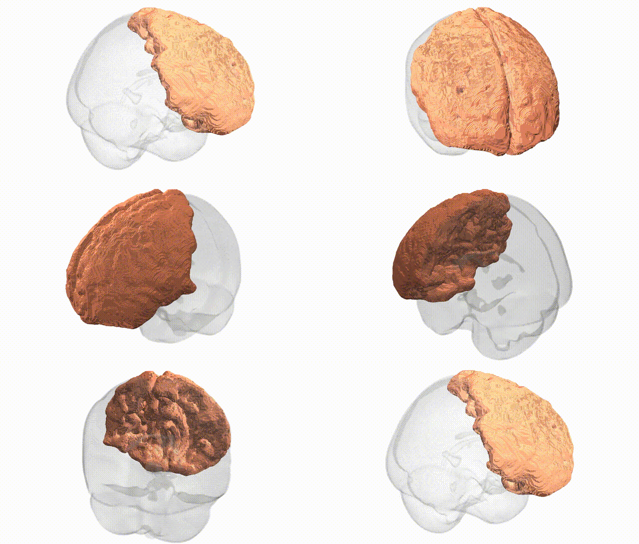
Full Quality Version: Download MP4
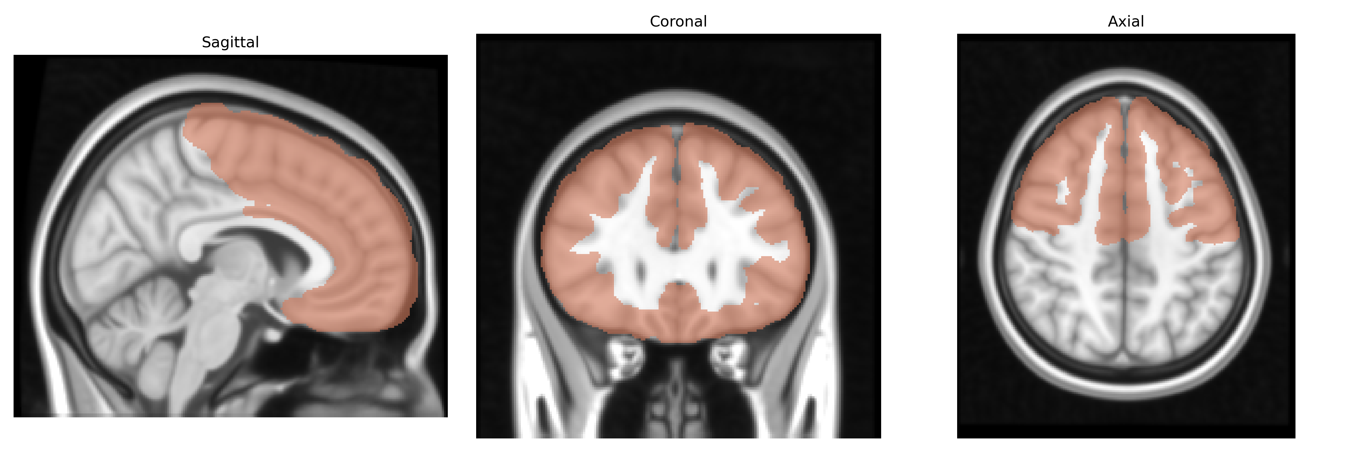
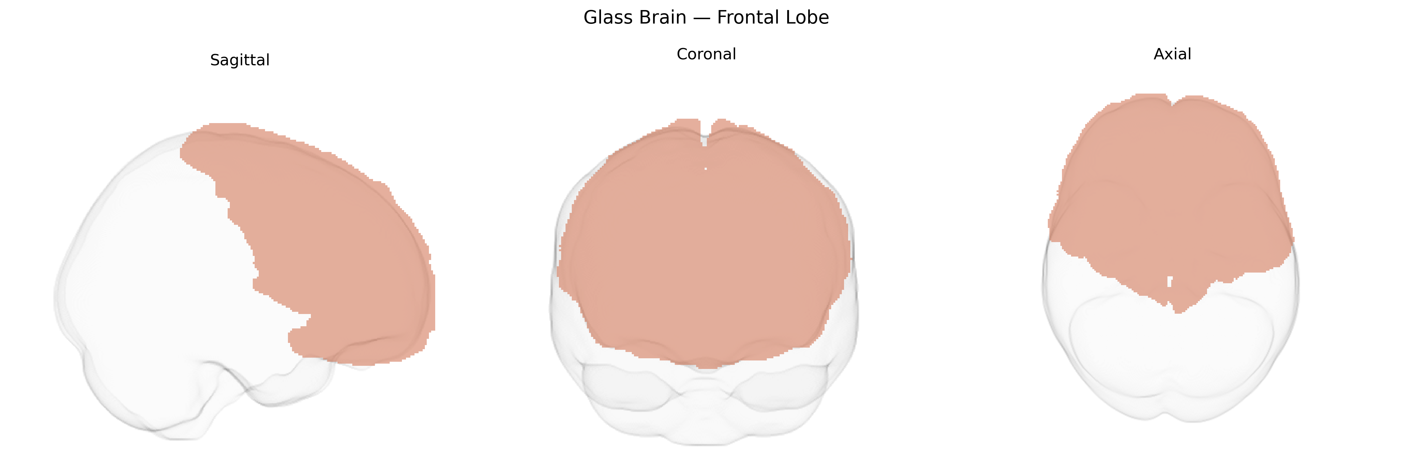
Wali Sidiqyar*, Gaurav Rudravaram*, Elyssa M. McMaster, Trent M. Schwartz, Adam M. Saunders, Kurt G. Schilling, Bennett A. Landman "Introducing SPINS: A Shared Public Visualization Library of Neuroanatomical Structures." Medical Imaging with Deep Learning- short paper
This resource is licensed under CC0 1.0 Universal (Public Domain).