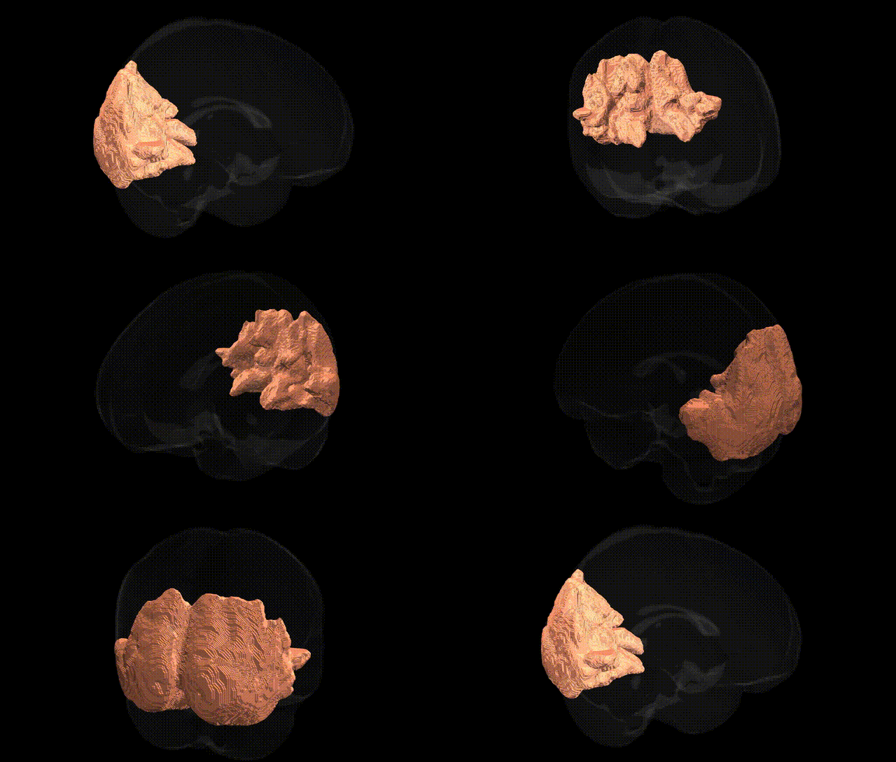
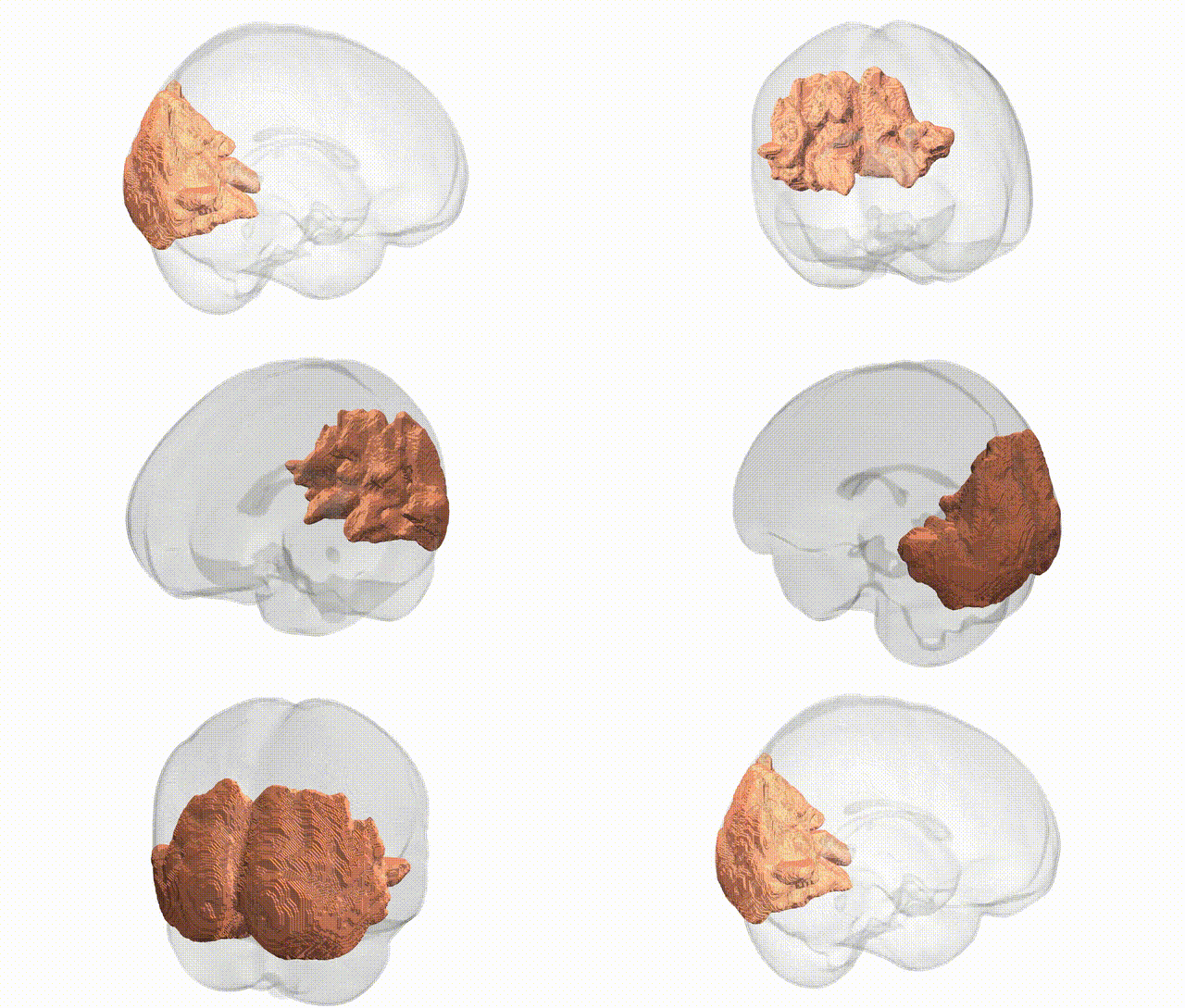
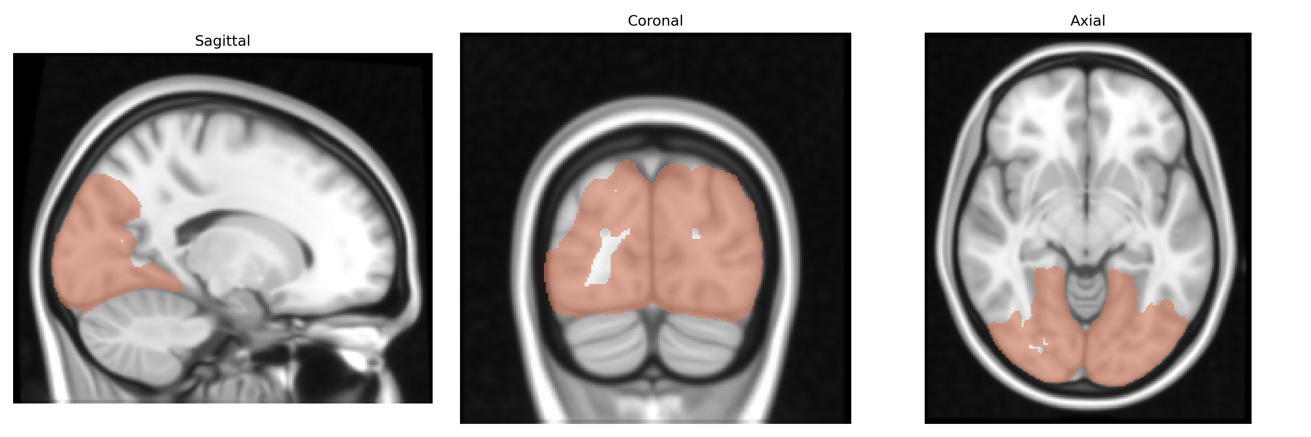
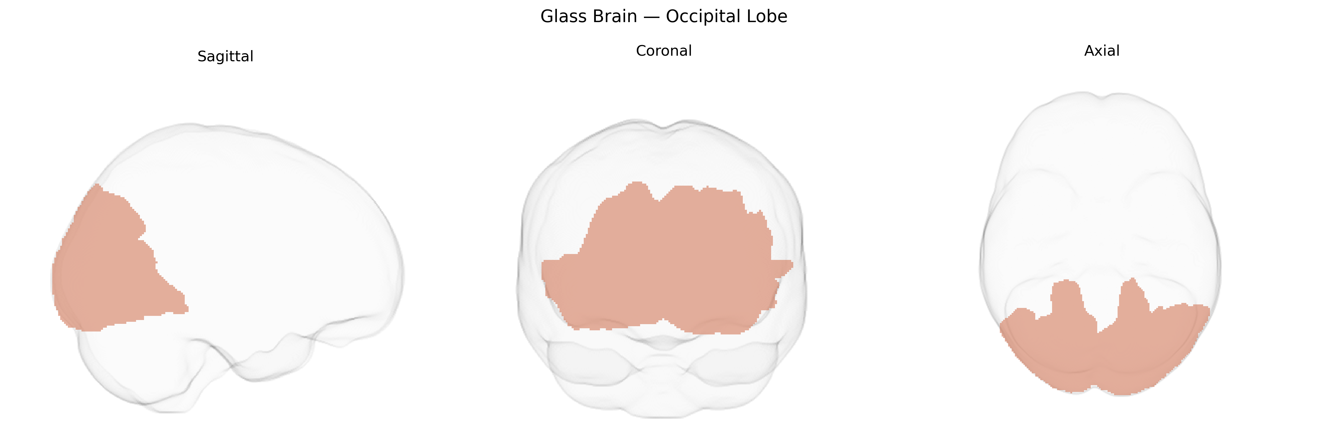
The bilateral occipital lobes are the paired posterior regions of the cerebral hemispheres primarily responsible for visual processing, encompassing the primary visual cortex (V1) and multiple higher-order visual areas involved in the analysis of orientation, motion, color, form, and spatial relationships. They receive the majority of their input from the lateral geniculate nucleus of the thalamus via the optic radiations and are organized retinotopically, with a systematic mapping of the visual field onto cortical surface. Damage to these regions can produce a range of visual disturbances, including cortical blindness, visual field defects, visual agnosias, and impairments in visuospatial perception. The occipital lobes interact extensively with parietal and temporal regions in dorsal and ventral visual streams to support visually guided actions and object recognition. Occipital lobe
Genetic associations involving the bilateral occipital lobe, as defined in the MNI Structural maxprob thr25 1 mm atlas, arise largely from imaging–genetics and GWAS of cortical structure, visual function, and neuropsychiatric or neurodevelopmental disorders. Variants in genes regulating neurodevelopment and axon guidance (for example, MAPK and Wnt pathway genes, cell-adhesion molecules such as those in the cadherin family, and synaptic genes including those in glutamatergic signaling) have been linked to occipital cortical thickness, surface area, and volume in large MRI–GWAS consortia such as ENIGMA and UK Biobank, with regionally specific hits often implicating loci near genes like RSPO3, TBR1, and others involved in cortical patterning. Occipital structural and functional variation shows polygenic overlap with disorders that affect visual processing or visual networks, including schizophrenia, major depression, autism spectrum disorder, and migraine, and with traits such as educational attainment, intelligence, and visual acuity, reflecting pleiotropic effects on both basic sensory cortices and higher-order visuocognitive systems. GWAS of occipital lobe epilepsy and photosensitive epilepsy have identified risk loci near genes involved in synaptic excitability and inhibitory–excitatory balance (for example, CACNA1A and GABRG2 in broader epileptic phenotypes), while rare variant and monogenic studies in disorders featuring occipital malformations or cortical blindness (such as some forms of periventricular heterotopia or posterior cortical atrophy) highlight roles for genes governing neuronal migration and cortical lamination. Overall, genetic influences on the bilateral occipital lobe appear highly polygenic and shared with other cortical regions, but with some enrichment of neurodevelopmental and visual-system genes that shape primary and associative visual cortex structure and function.
Overview generated by GPT-4o (2026).
Region ID: 5
Hemisphere: bilateral
Atlas: MNI Structural maxprob thr25 1mm
Full Quality Version: Download MP4
Full Quality Version: Download MP4
Wali Sidiqyar*, Gaurav Rudravaram*, Elyssa M. McMaster, Trent M. Schwartz, Adam M. Saunders, Kurt G. Schilling, Bennett A. Landman "Introducing SPINS: A Shared Public Visualization Library of Neuroanatomical Structures." Medical Imaging with Deep Learning- short paper
This resource is licensed under CC0 1.0 Universal (Public Domain).