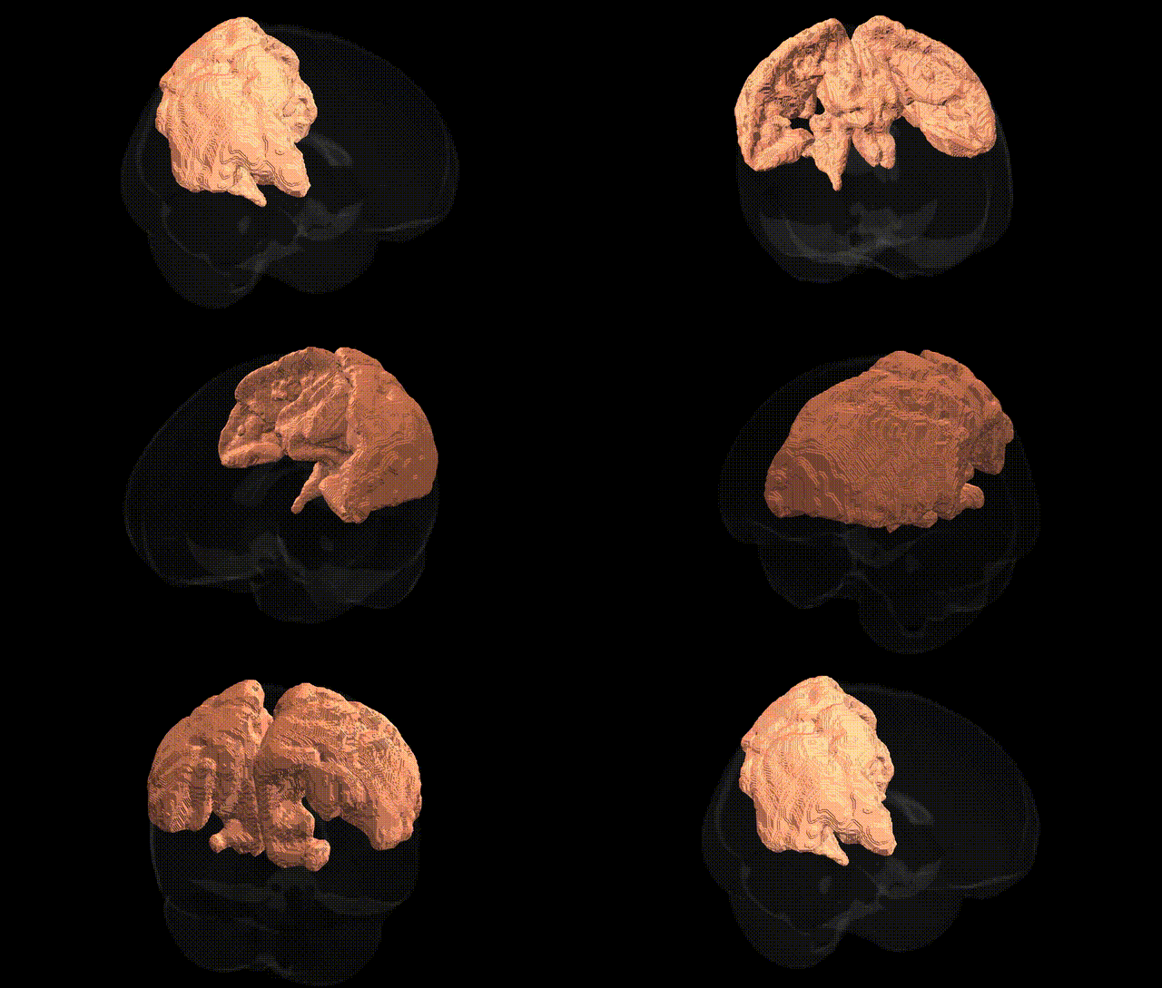
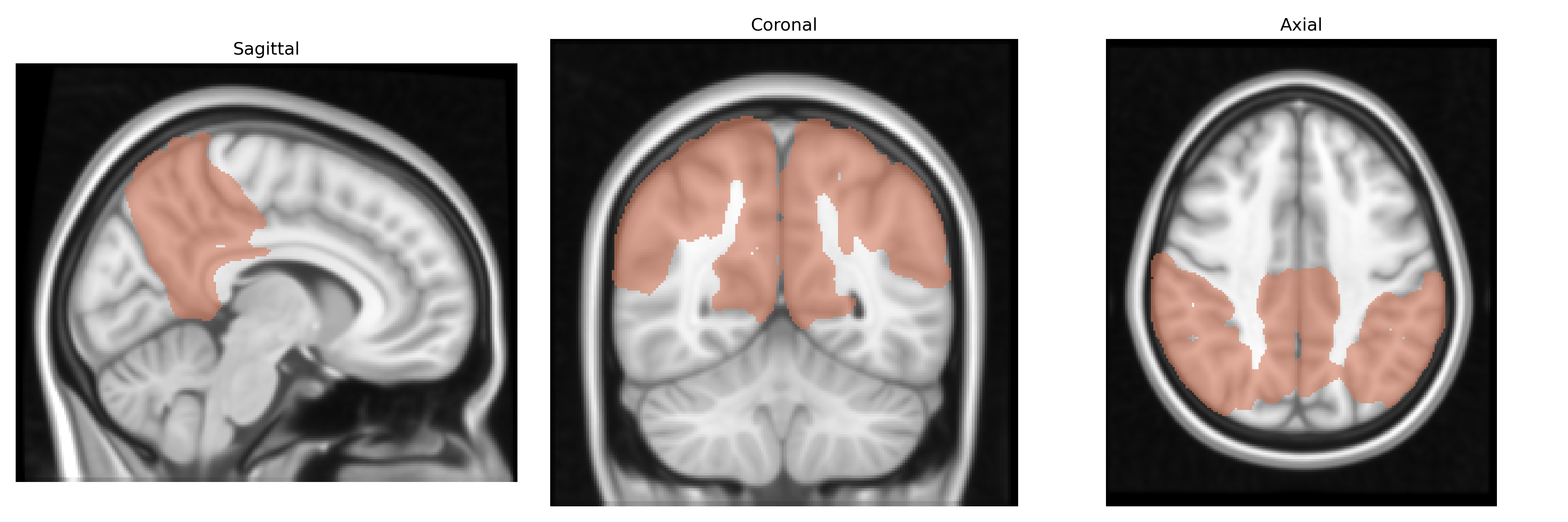
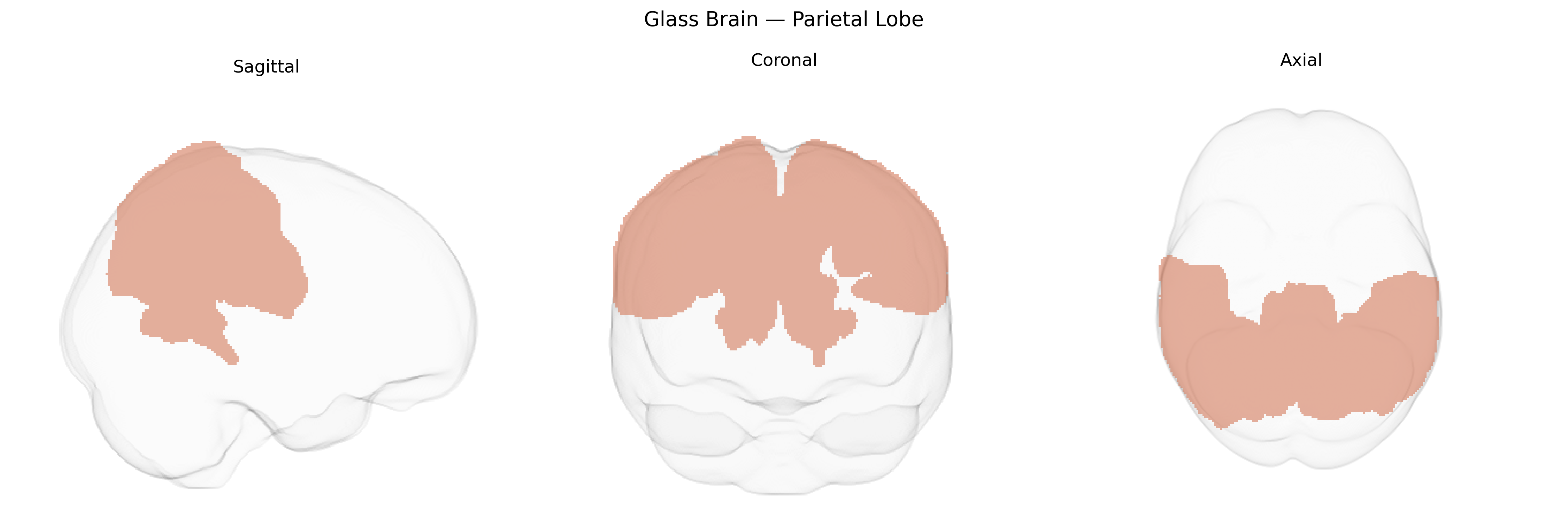
The bilateral parietal lobe, as defined in the MNI Structural maxprob thr25 1 mm atlas, encompasses cortical territories in both hemispheres located posterior to the central sulcus and superior to the lateral (Sylvian) fissure, including superior and inferior parietal lobules and adjoining regions bordering frontal, temporal, and occipital cortices. This region participates in multimodal sensory integration by combining somatosensory, visual, and auditory information to support spatial perception, body schema, and goal-directed actions such as reaching, grasping, and visuomotor coordination. It is critically involved in attentional allocation, numerical and symbolic processing, and aspects of language, particularly in mapping sensory inputs to motor outputs and in higher-order cognitive operations requiring spatial transformation. Lesions in bilateral or dominant parietal regions can lead to syndromes such as hemispatial neglect, apraxia, and Gerstmann’s syndrome, reflecting its central role in integrating sensory information with motor and cognitive functions. Parietal lobe
The bilateral parietal lobes, encompassing regions such as the superior and inferior parietal cortex as defined in the MNI Structural maxprob thr25 1mm Atlas, have been repeatedly implicated in genetic studies of cognition, neurodevelopmental and neuropsychiatric disorders, and brain structure. GWAS of cortical thickness and surface area have identified common variants in genes including KIAA0586, CENPW, TBR1, and regulatory loci near WNT and FGF pathway genes that influence parietal morphology and maturation, often overlapping with loci associated with general intelligence, educational attainment, and executive function. Parietal regions show strong heritability for both structure and functional activation, and imaging-genetics work has linked schizophrenia-, bipolar-, and major depression–associated loci (for example in CACNA1C, ZNF804A, and GRM3) to altered parietal connectivity and activation during working memory and attention tasks. ADHD GWAS and polygenic risk studies report associations between risk variants (e.g., in FOXP2, SLC6A3, and dopaminergic signaling genes) and atypical parietal activation or structure underlying attentional control, while autism spectrum disorder risk genes involved in synapse formation and axon guidance (such as CNTNAP2 and NRXN1) have been associated with atypical parietal connectivity supporting social cognition and multisensory integration. In addition, Alzheimer’s disease and other dementias show genetic links to parietal atrophy and hypometabolism; APOE ε4, CLU, and BIN1 carriers exhibit accelerated parietal cortical thinning and posterior default-mode network disruption. Overall, GWAS and candidate-gene imaging-genetics studies converge on the parietal lobes as a genetically sensitive hub for attention, visuospatial processing, numerical cognition, and integration of multimodal information, mediating risk and resilience across multiple brain-related traits and disorders.
Overview generated by GPT-4o (2026).
Region ID: 6
Hemisphere: bilateral
Atlas: MNI Structural maxprob thr25 1mm
Full Quality Version: Download MP4
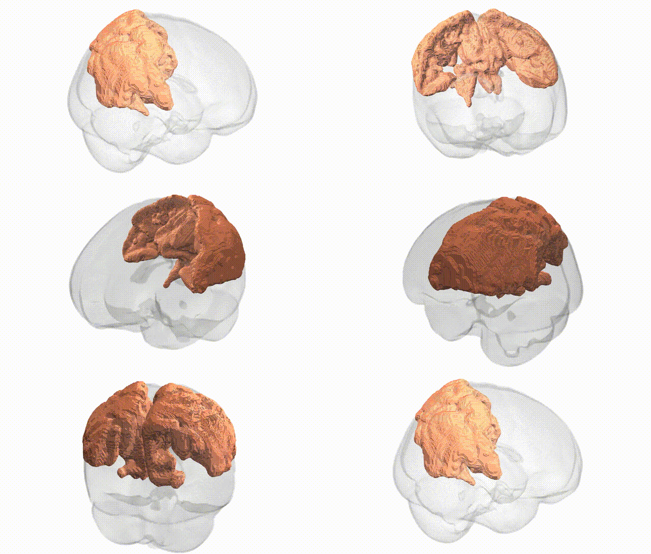
Full Quality Version: Download MP4
Wali Sidiqyar*, Gaurav Rudravaram*, Elyssa M. McMaster, Trent M. Schwartz, Adam M. Saunders, Kurt G. Schilling, Bennett A. Landman "Introducing SPINS: A Shared Public Visualization Library of Neuroanatomical Structures." Medical Imaging with Deep Learning- short paper
This resource is licensed under CC0 1.0 Universal (Public Domain).