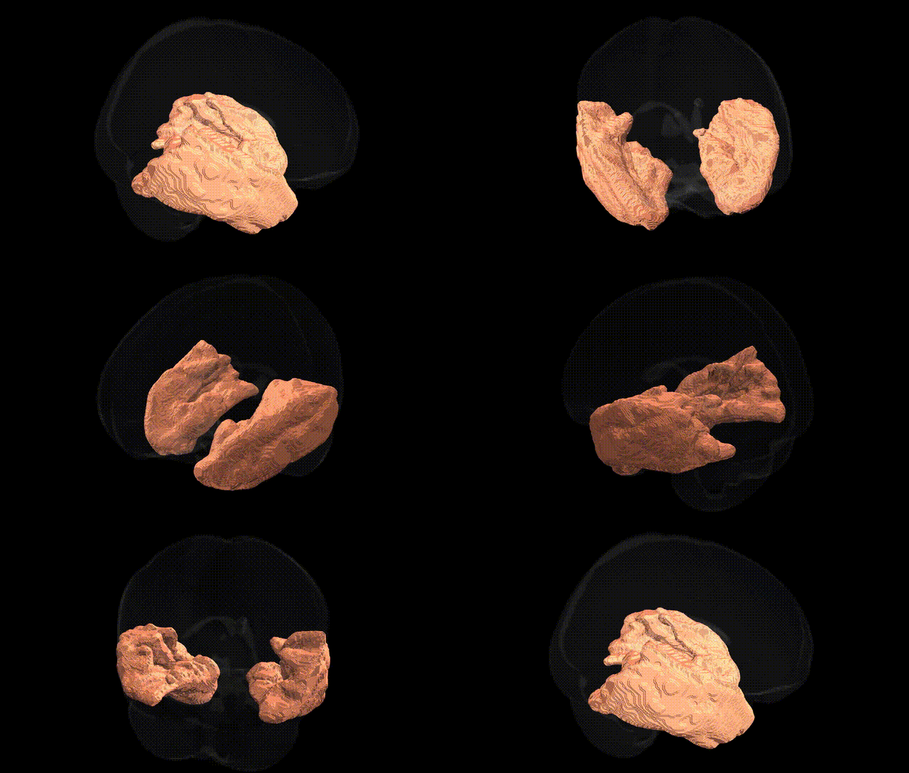
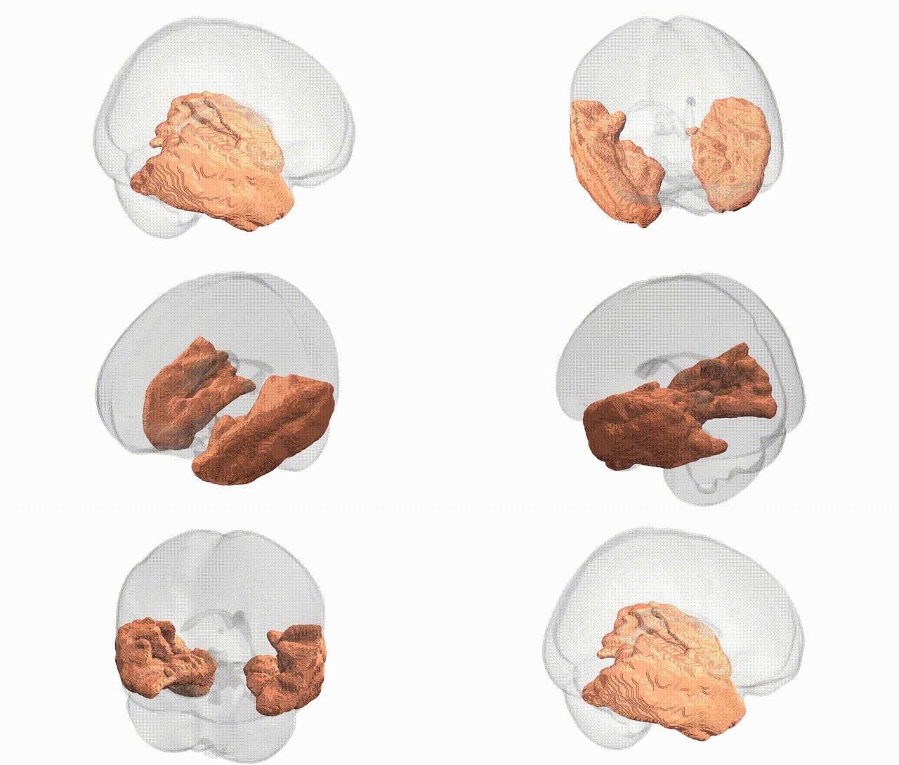
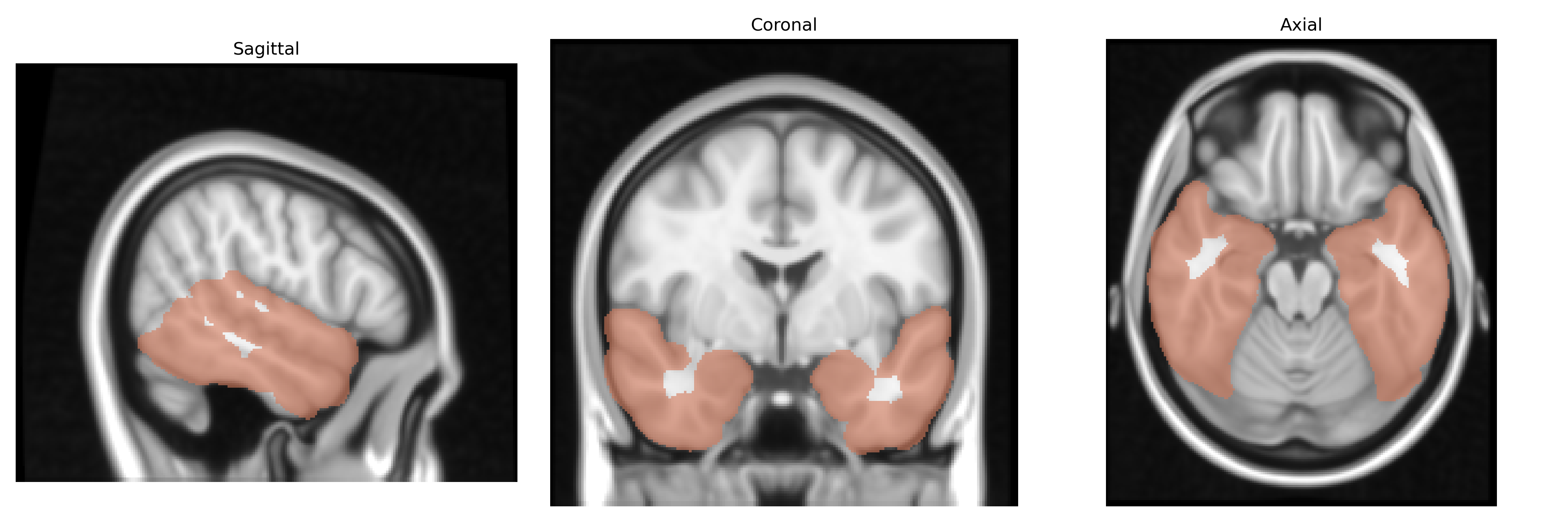
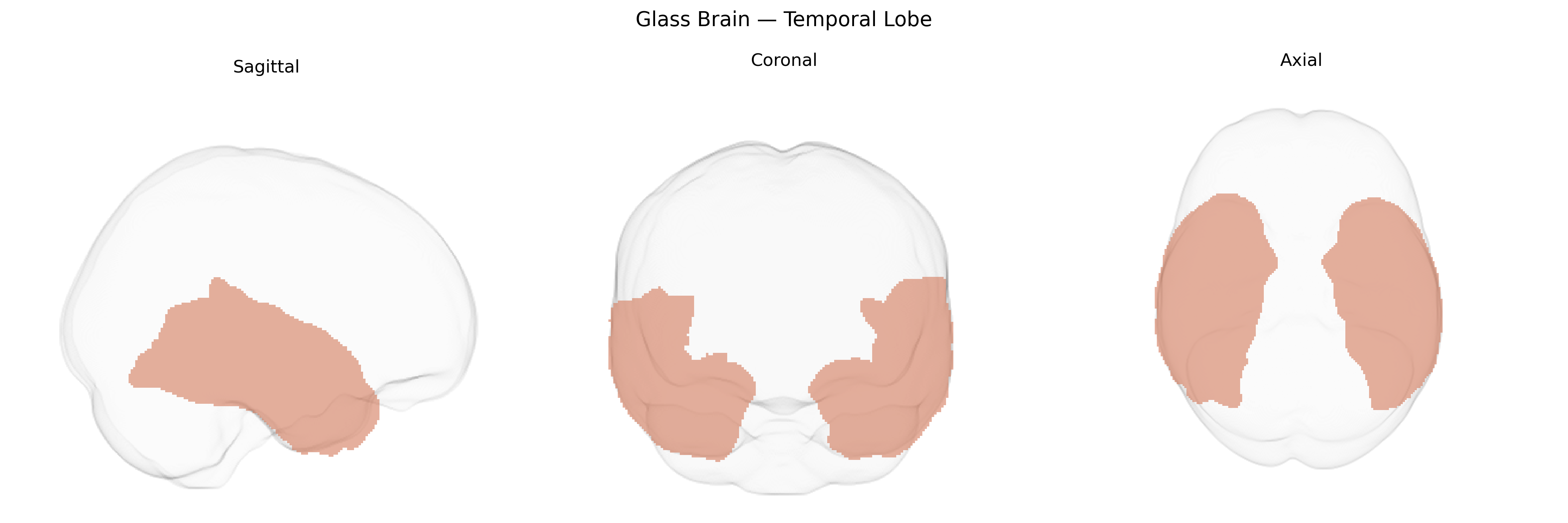
The bilateral temporal lobes comprise paired cortical regions located on the lateral aspects of the cerebral hemispheres, inferior to the lateral (Sylvian) fissure and anterior to the occipital lobes, playing central roles in auditory processing, language comprehension, semantic memory, and aspects of social and emotional cognition. Structurally, they include the superior, middle, and inferior temporal gyri, as well as medial temporal structures such as the hippocampus and amygdala, interconnected with frontal and parietal association cortices via long-range white matter tracts (e.g., arcuate fasciculus, uncinate fasciculus). Functionally, the superior temporal cortex is critical for primary and higher-order auditory perception, Wernicke’s area in the dominant hemisphere supports language comprehension, and anterior/mid temporal cortices contribute to object recognition, semantic processing, and autobiographical memory. Medial temporal components mediate episodic memory encoding and retrieval, while limbic-related regions contribute to emotional learning and affective evaluation of sensory stimuli.
The bilateral temporal lobes, encompassing structures such as the superior, middle, and inferior temporal gyri, temporal poles, and medial temporal regions, show convergent genetic associations from GWAS and imaging genetics studies, particularly those using voxel- and region-based measures aligned to MNI atlases. Common variants in genes like APOE, CLU, PICALM, BIN1, ABCA7, and CR1 are robustly associated with temporal lobe atrophy and cortical thinning in Alzheimer’s disease, while TOMM40 and SORL1 influence medial and lateral temporal volumes, especially in aging cohorts. Variants near GRIN2A, GRIN2B, BDNF, and NRGN have been linked to temporal lobe cortical thickness and surface area in large consortia such as ENIGMA, and polygenic risk scores for schizophrenia, bipolar disorder, and major depression correlate with structural alterations in temporal regions involved in language, auditory processing, and social cognition. GWAS of temporal lobe epilepsy implicate loci in SCN1A, SCN2A, GABRA2, and other ion channel and synaptic genes, aligning with hippocampal and parahippocampal involvement, while autism-spectrum and language-impairment studies highlight variants in CNTNAP2, FOXP2, and DCDC2 that modulate temporal lobe structure and connectivity. More broadly, multivariate neuroimaging GWAS demonstrate that genetic factors influencing general cognitive ability, educational attainment, and reading/language skills exert significant effects on temporal lobe morphology, suggesting this bilateral region is a key anatomical target of diverse neuropsychiatric and cognitive genetic architectures.
Overview generated by GPT-4o (2026).
Region ID: 8
Hemisphere: bilateral
Atlas: MNI Structural maxprob thr25 1mm
Full Quality Version: Download MP4
Full Quality Version: Download MP4
Wali Sidiqyar*, Gaurav Rudravaram*, Elyssa M. McMaster, Trent M. Schwartz, Adam M. Saunders, Kurt G. Schilling, Bennett A. Landman "Introducing SPINS: A Shared Public Visualization Library of Neuroanatomical Structures." Medical Imaging with Deep Learning- short paper
This resource is licensed under CC0 1.0 Universal (Public Domain).