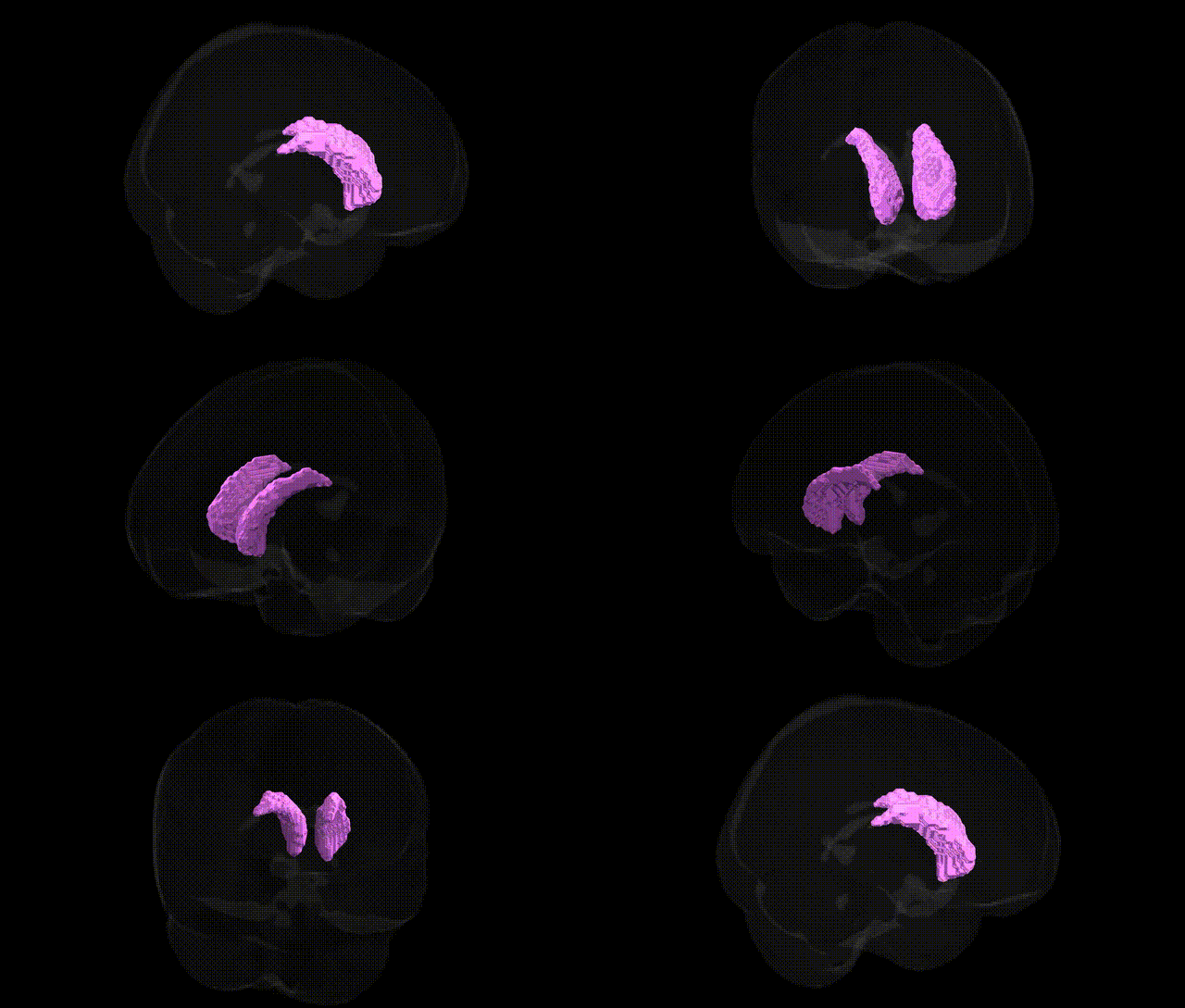
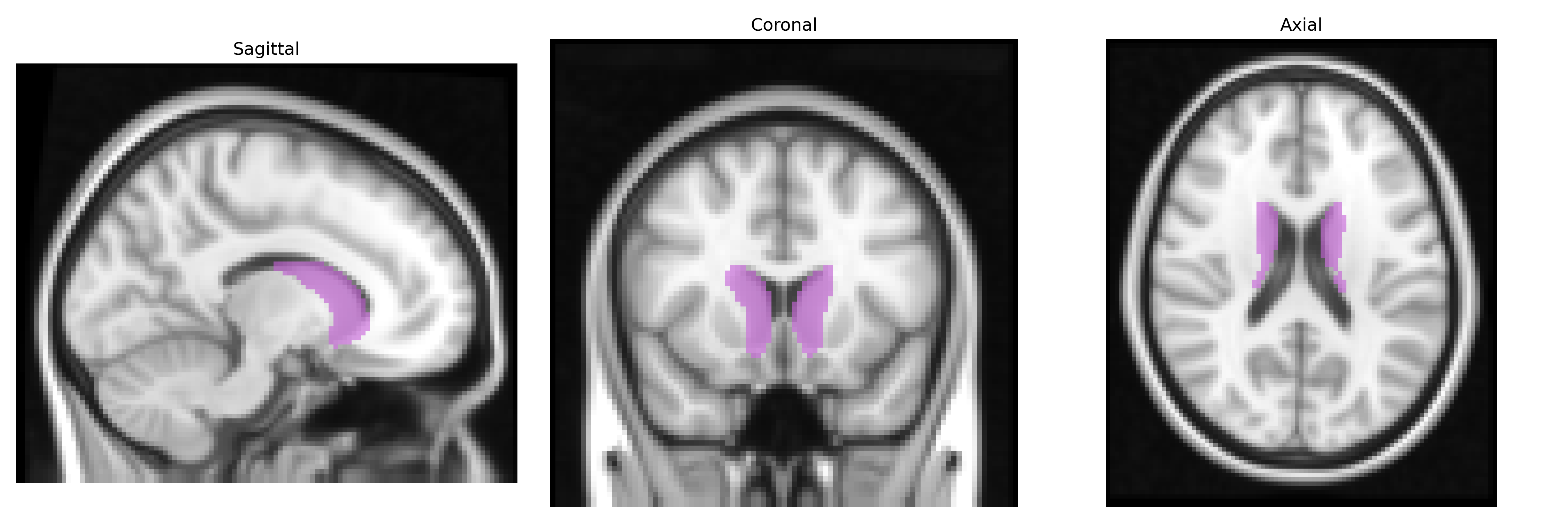
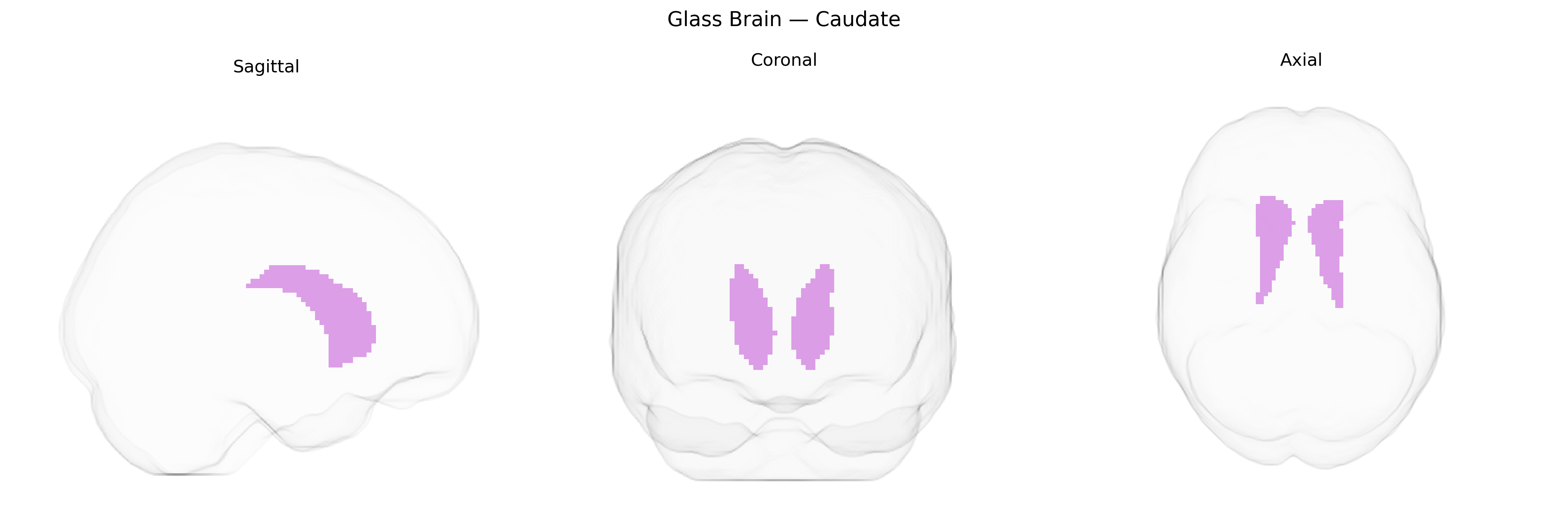
The bilateral caudate, as defined in the MNI Structural maxprob thr25 2 mm Atlas, comprises the paired caudate nuclei situated in the dorsal medial aspect of the telencephalon, forming a major component of the dorsal striatum within the basal ganglia circuitry. Each caudate nucleus is an elongated C-shaped gray-matter structure with head, body, and tail portions following the lateral ventricle, and is heavily interconnected with the cortex, thalamus, and other basal ganglia nuclei. Functionally, the caudate is involved in motor control, action selection, goal-directed behavior, habit formation, learning, and aspects of cognition and emotion, integrating cortical inputs (particularly from prefrontal and associative cortices) and relaying them via the basal ganglia-thalamo-cortical loops. A related article is Caudate nucleus.
The bilateral caudate nucleus, as defined in the MNI Structural maxprob thr25 2 mm atlas, shows robust heritability and has been implicated in multiple genome-wide association studies (GWAS) linking its volume, shape, and connectivity to variants in genes involved in neurodevelopment, synaptic function, and dopaminergic signaling, such as DRD2, BDNF, CNTNAP2, and several loci near HMGA2, KTN1, and FAT3 identified by large imaging-genetics consortia (e.g., ENIGMA, UK Biobank). Common variants influencing caudate structure and function have been associated with a range of neuropsychiatric and neurodegenerative disorders, including schizophrenia, bipolar disorder, major depressive disorder, obsessive–compulsive disorder, attention-deficit/hyperactivity disorder, Tourette syndrome, and Parkinson’s and Huntington’s diseases, as well as cognitive traits such as general intelligence, executive function, and response inhibition. Polygenic risk scores for these conditions often correlate with caudate volume or activity, and several risk genes for frontostriatal circuit dysfunction (for example, CACNA1C, GRIN2A, and genes involved in glutamatergic and GABAergic transmission) show convergent effects on caudate-related networks, underscoring this region’s role as a genetically influenced hub in corticostriatal pathways that contribute to both normal individual differences and vulnerability to psychiatric and movement disorders.
Overview generated by GPT-4o (2026).
Region ID: 1
Hemisphere: bilateral
Atlas: MNI Structural maxprob thr25 2mm
Full Quality Version: Download MP4
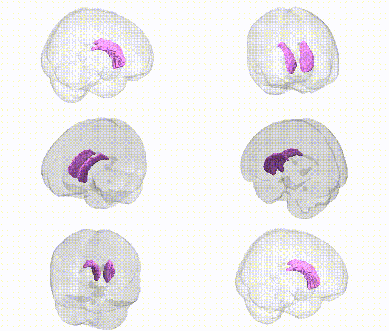
Full Quality Version: Download MP4
Wali Sidiqyar*, Gaurav Rudravaram*, Elyssa M. McMaster, Trent M. Schwartz, Adam M. Saunders, Kurt G. Schilling, Bennett A. Landman "Introducing SPINS: A Shared Public Visualization Library of Neuroanatomical Structures." Medical Imaging with Deep Learning- short paper
This resource is licensed under CC0 1.0 Universal (Public Domain).