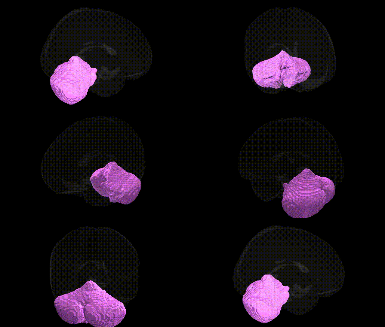
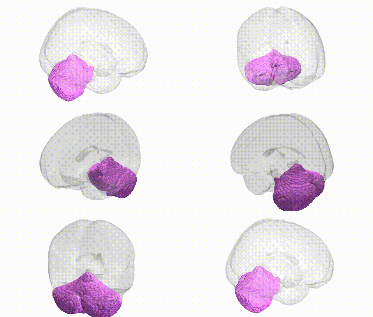
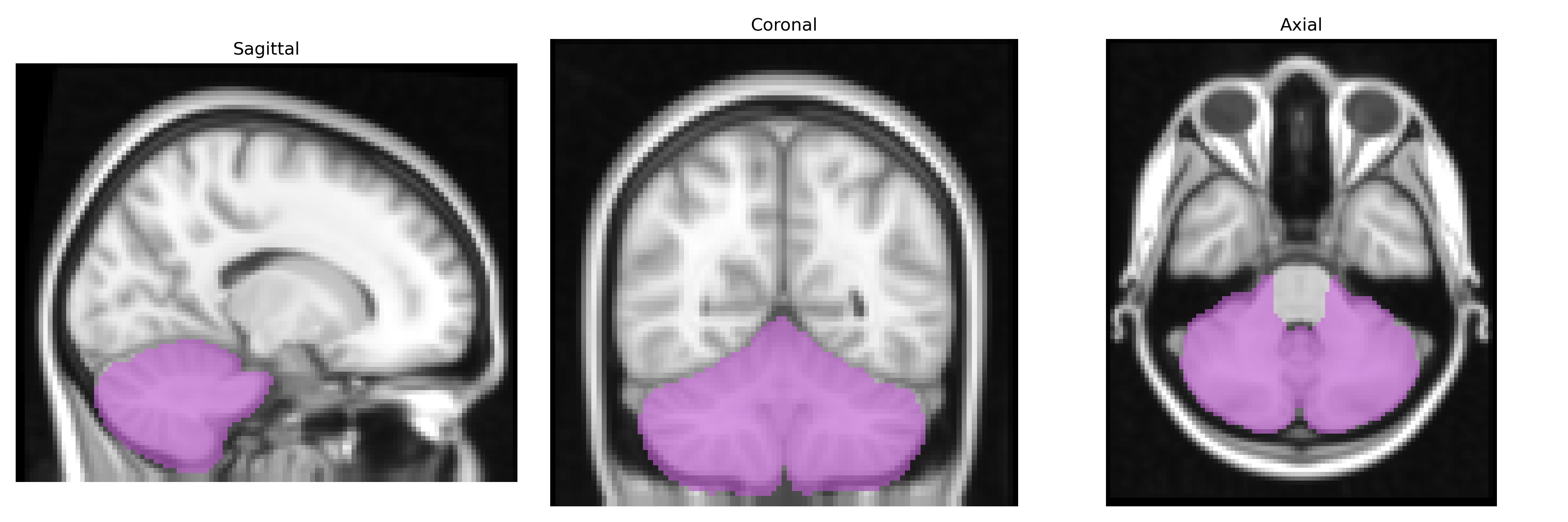
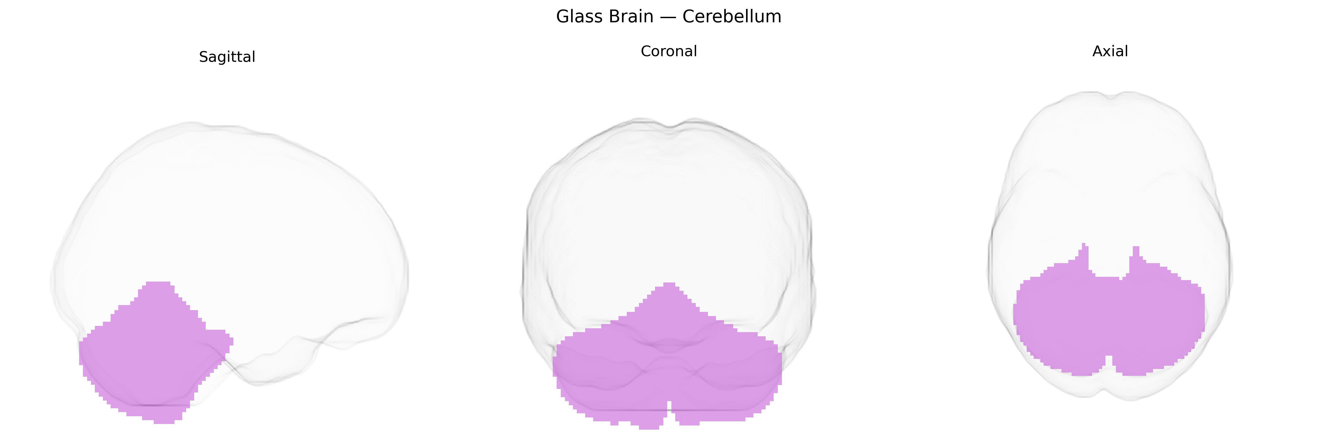
The bilateral cerebellum, as defined in the MNI Structural maxprob thr25 2 mm atlas, comprises the paired cerebellar hemispheres located in the posterior cranial fossa beneath the occipital lobes and dorsal to the brainstem. Structurally, it is organized into a midline vermis and lateral hemispheres, with a highly folded cortical surface overlying deep white matter and the dentate and other cerebellar nuclei. Functionally, the bilateral cerebellum is critical for the coordination, timing, and precision of voluntary movements, maintenance of posture and balance, and fine-tuning of motor commands through integration of proprioceptive, vestibular, and cortical inputs. Increasing evidence also implicates the cerebellar hemispheres in cognitive and affective processes via cerebrocerebellar loops connecting with association cortices and limbic structures.
Cerebellum
The bilateral cerebellum, as defined in the MNI Structural maxprob thr25 2mm atlas, has been implicated in multiple genetic and genome-wide association studies (GWAS), reflecting its role beyond motor coordination in cognition, affect, and neurodevelopmental processes. Large neuroimaging GWAS (e.g., ENIGMA, UK Biobank) have identified common variants influencing cerebellar volume and morphology, including loci near genes such as KATNAL2, MAPT, NKX6-2, and FOXP1, which are broadly involved in neurodevelopment, synaptic function, or axonal processes. Polygenic overlap has been observed between cerebellar structural measures and psychiatric traits—most consistently with schizophrenia, bipolar disorder, major depressive disorder, and autism spectrum disorder—where risk variants often map to pathways regulating synaptic plasticity, glutamatergic signaling, and neuronal migration, all critical to cerebellar circuit formation. Cerebellar GWAS signals also show genetic correlation with cognitive performance, educational attainment, and general intelligence, suggesting shared genetic architecture between cerebellar anatomy and higher-order cognition. In neurodegenerative disease, variants in genes such as ATXN1–3, CACNA1A, and others causing spinocerebellar ataxias, as well as C9orf72 and MAPT in frontotemporal dementia and parkinsonian syndromes, highlight monogenic and polygenic contributions to cerebellar vulnerability. Additionally, imaging-genetics work has linked risk alleles for ADHD, anxiety, and substance use traits to altered cerebellar activation and connectivity, reinforcing the view that cerebellar structure and function are shaped by distributed neurodevelopmental genetic effects that contribute to a wide spectrum of behavioral and clinical phenotypes.
Overview generated by GPT-4o (2026).
Region ID: 2
Hemisphere: bilateral
Atlas: MNI Structural maxprob thr25 2mm
Full Quality Version: Download MP4
Full Quality Version: Download MP4
Wali Sidiqyar*, Gaurav Rudravaram*, Elyssa M. McMaster, Trent M. Schwartz, Adam M. Saunders, Kurt G. Schilling, Bennett A. Landman "Introducing SPINS: A Shared Public Visualization Library of Neuroanatomical Structures." Medical Imaging with Deep Learning- short paper
This resource is licensed under CC0 1.0 Universal (Public Domain).