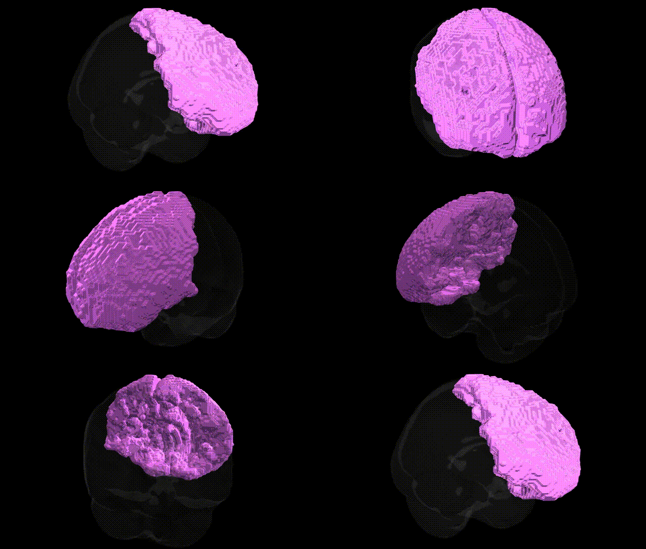
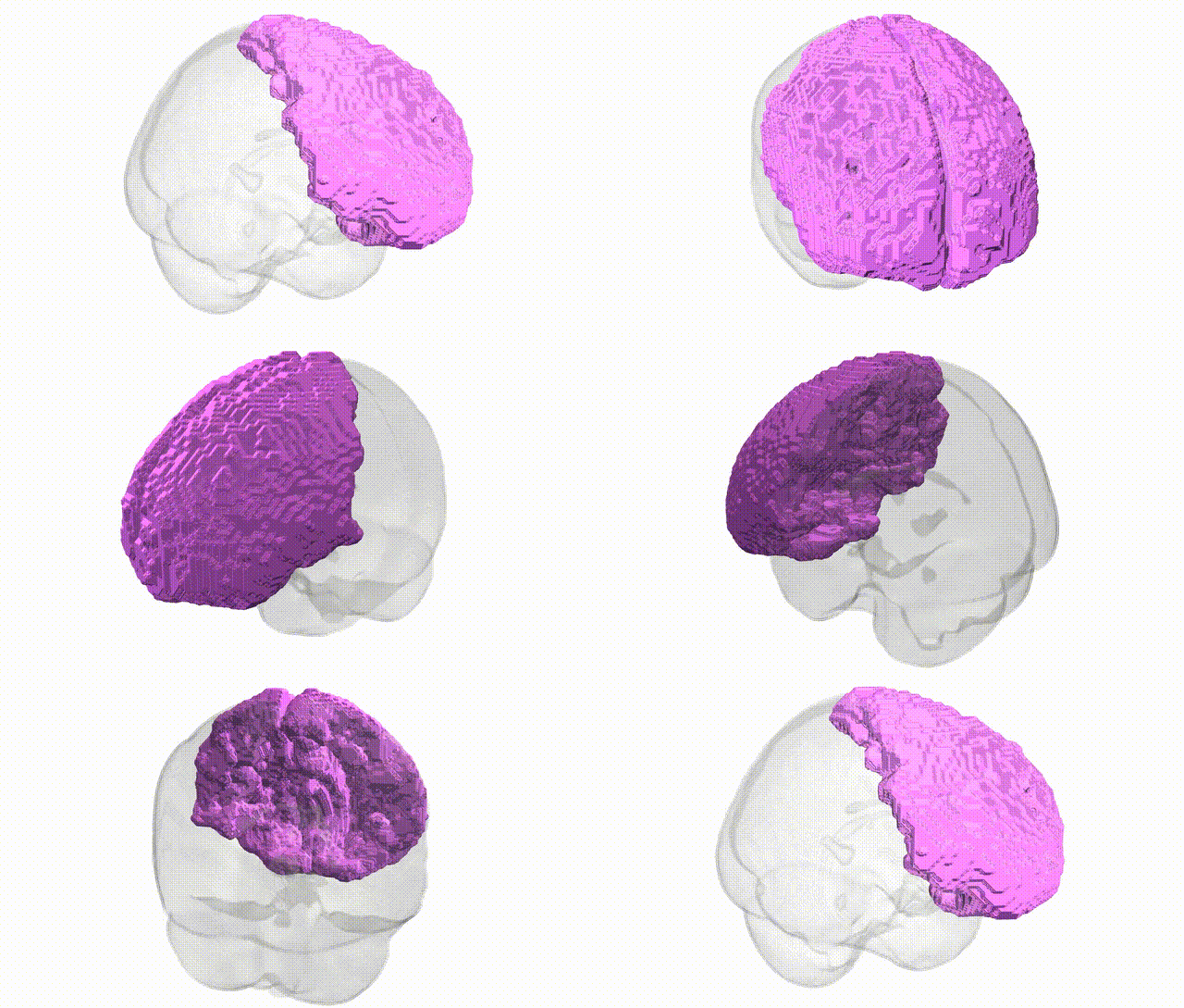
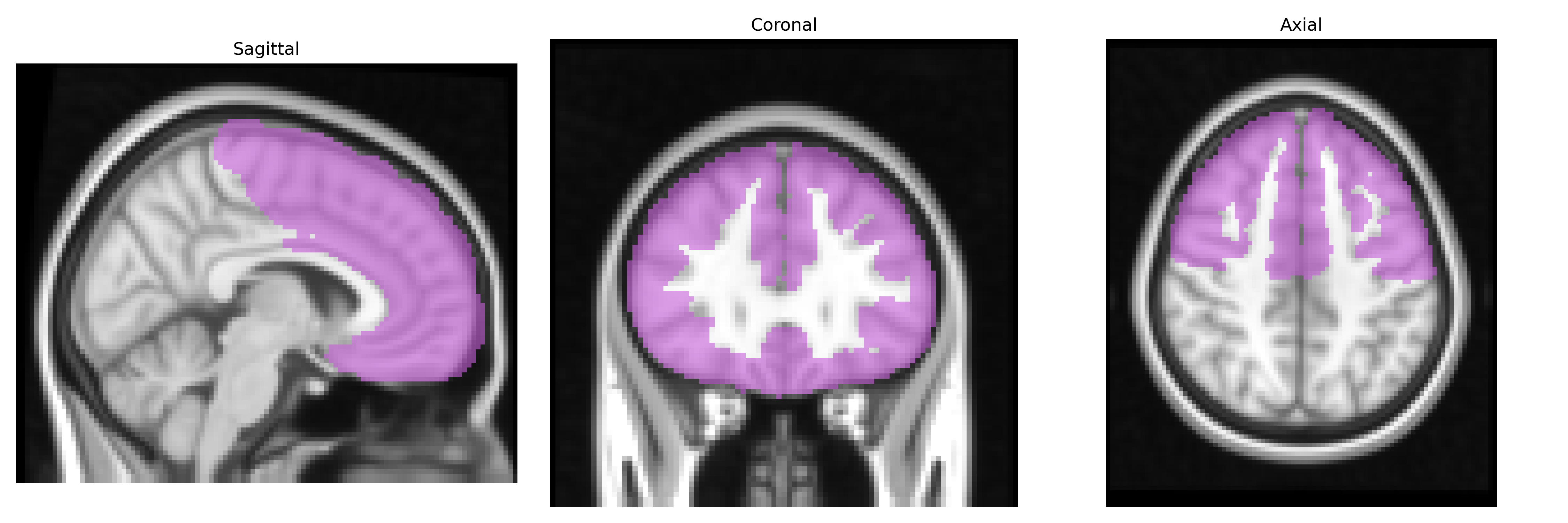
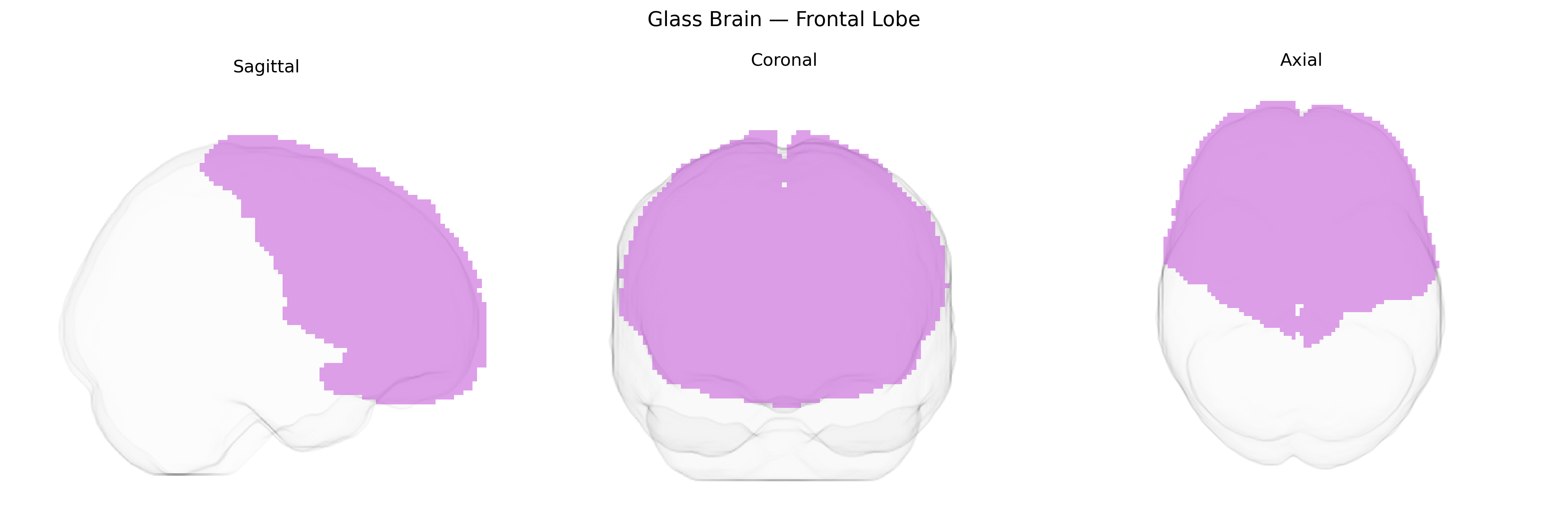
The bilateral frontal lobe, as defined in the MNI Structural maxprob thr25 2 mm Atlas, comprises the paired cortical territories located anterior to the central sulcus that subserve a broad range of higher-order cognitive, motor, and affective functions. This region includes primary and premotor areas involved in voluntary movement planning and execution, as well as prefrontal territories critical for executive functions such as working memory, decision-making, planning, inhibitory control, and social cognition. Frontal lobe networks integrate multimodal sensory input with internal goals and reward signals to guide behavior, support language production via connections with perisylvian regions, and regulate emotional responses through extensive reciprocal connections with limbic and subcortical structures. Developmentally and clinically, the frontal lobes are central to the maturation of goal-directed behavior and are particularly vulnerable in traumatic, neurodegenerative, and psychiatric conditions that impair executive functioning and personality. Frontal lobe
The bilateral frontal lobes, as delineated in the MNI Structural maxprob thr25 2mm atlas, are a central substrate for complex cognition, executive function, and emotion regulation, and genetic studies have repeatedly implicated variants affecting this region in a wide range of traits and disorders. Large neuroimaging GWAS consortia (e.g., ENIGMA, UK Biobank) have identified common variants in genes such as HMGA2, IGF1, MAPT, and those in neurodevelopmental and synaptic pathways (e.g., CNTNAP2, DISC1, GRIN2B) that are associated with frontal lobe volume, cortical thickness, and surface area. Polygenic risk scores for schizophrenia, bipolar disorder, major depressive disorder, ADHD, and autism spectrum disorder show robust associations with structural and functional alterations in bilateral frontal regions, particularly dorsolateral and orbitofrontal subregions. Genes involved in neuroplasticity and neurotransmission (including BDNF, COMT, and dopaminergic and glutamatergic receptor genes) have been linked to frontal lobe–dependent traits such as intelligence, working memory, impulsivity, risk-taking, and personality dimensions (e.g., neuroticism and extraversion), often via GWAS that relate genotype to MRI-derived measures of frontal structure or activity. Additionally, APOE and other loci influencing vascular and neurodegenerative processes show associations with age-related frontal atrophy and cognitive decline, underscoring the frontal lobes as a convergent target of diverse genetic influences on brain development, psychiatric vulnerability, and cognitive performance.
Overview generated by GPT-4o (2026).
Region ID: 3
Hemisphere: bilateral
Atlas: MNI Structural maxprob thr25 2mm
Full Quality Version: Download MP4
Full Quality Version: Download MP4
Wali Sidiqyar*, Gaurav Rudravaram*, Elyssa M. McMaster, Trent M. Schwartz, Adam M. Saunders, Kurt G. Schilling, Bennett A. Landman "Introducing SPINS: A Shared Public Visualization Library of Neuroanatomical Structures." Medical Imaging with Deep Learning- short paper
This resource is licensed under CC0 1.0 Universal (Public Domain).