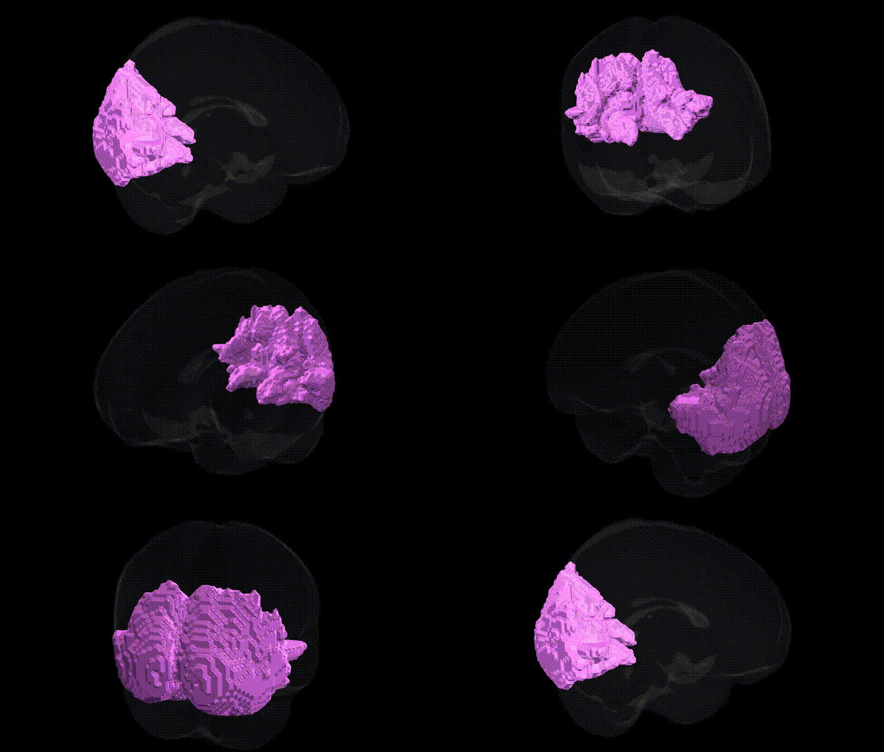
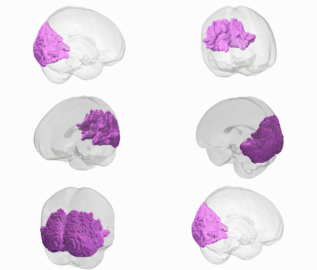
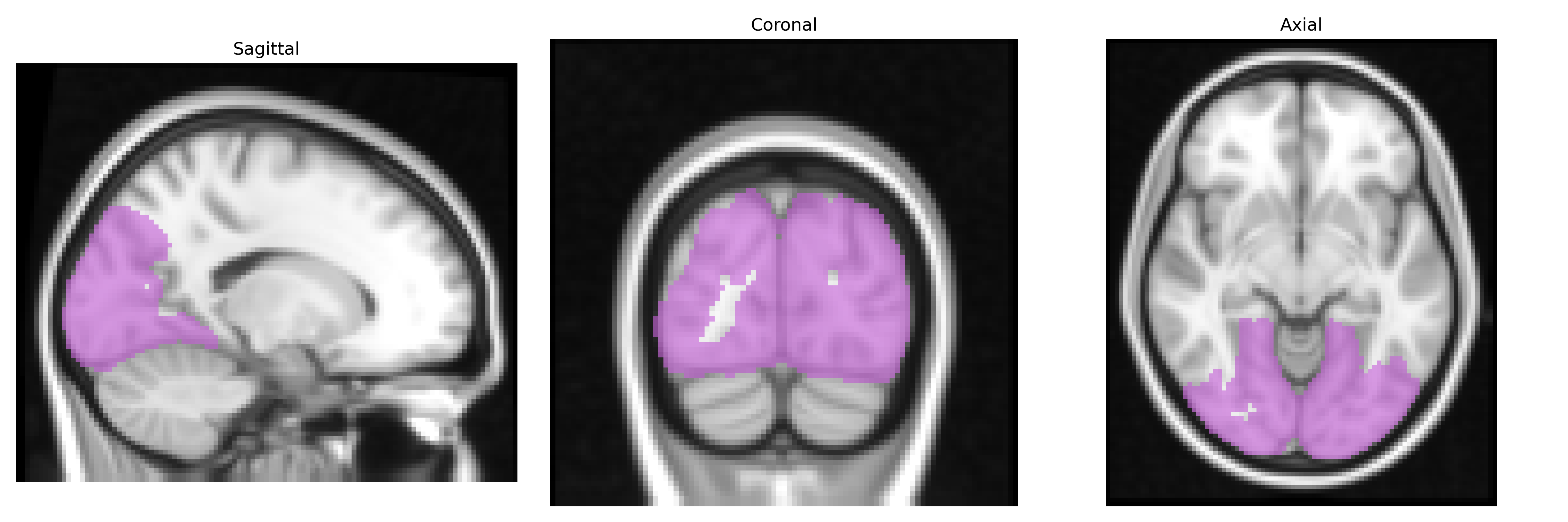
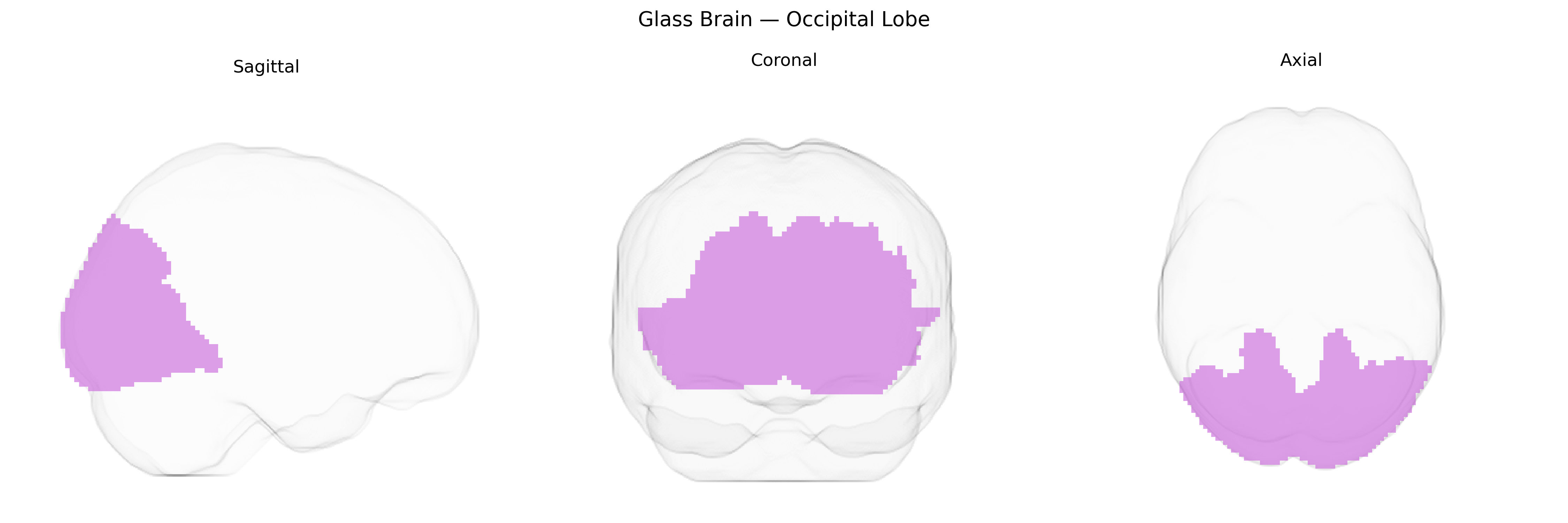
The bilateral occipital lobe, as defined in the MNI Structural maxprob thr25 2mm Atlas, comprises the paired cortical regions located at the posterior poles of the cerebral hemispheres, primarily responsible for processing visual information. This lobe contains the primary visual cortex (V1, Brodmann area 17) and multiple secondary visual areas (e.g., BA 18, 19) that support functions such as basic visual feature detection, motion perception, color processing, form recognition, and visual spatial integration. The occipital lobe receives major input via the geniculostriate pathway from the lateral geniculate nucleus of the thalamus and participates in extensive reciprocal connections with parietal and temporal cortices, forming the dorsal and ventral visual streams critical for visually guided actions and object recognition. Lesions in this bilateral region can lead to a range of visual deficits, including cortical blindness, visual field cuts, and higher-order visual agnosias. Occipital lobe
The bilateral occipital lobes, encompassing primary and secondary visual cortices in the MNI Structural maxprob thr25 2 mm atlas, have been implicated in numerous genetic studies examining visual processing, cortical structure, and neuropsychiatric traits. GWAS of brain imaging phenotypes (e.g., ENIGMA, UK Biobank) have linked common variants in genes related to neurodevelopment, synaptic function, and myelination (such as those near HMGA2, DCC, PAX6, and multiple loci on chromosomes 6, 10, and 17) to occipital cortical thickness, surface area, and volume. Variants associated with visual acuity and refractive error (e.g., near GJD2, RASGRF1) show downstream effects on occipital activation patterns in functional imaging studies, while genes involved in glutamatergic signaling and synaptic plasticity (including GRIN2B and BDNF) have been associated with individual differences in visual perception and occipital activity. Occipital lobe measures also appear as endophenotypes in GWAS of migraine, schizophrenia, bipolar disorder, and major depression, where risk loci overlap with regions influencing cortical morphology and connectivity, and in neurodevelopmental conditions such as autism spectrum disorder and ADHD, occipital structural and functional alterations co-occur with polygenic risk scores enriched for synaptic and neuronal differentiation pathways. Collectively, genetic associations suggest that common variation affecting neurodevelopmental patterning, excitatory–inhibitory balance, and visual system wiring contributes to occipital lobe structure and function, with downstream relevance to sensory processing, cognition, and vulnerability to several brain disorders.
Overview generated by GPT-4o (2026).
Region ID: 5
Hemisphere: bilateral
Atlas: MNI Structural maxprob thr25 2mm
Full Quality Version: Download MP4
Full Quality Version: Download MP4
Wali Sidiqyar*, Gaurav Rudravaram*, Elyssa M. McMaster, Trent M. Schwartz, Adam M. Saunders, Kurt G. Schilling, Bennett A. Landman "Introducing SPINS: A Shared Public Visualization Library of Neuroanatomical Structures." Medical Imaging with Deep Learning- short paper
This resource is licensed under CC0 1.0 Universal (Public Domain).