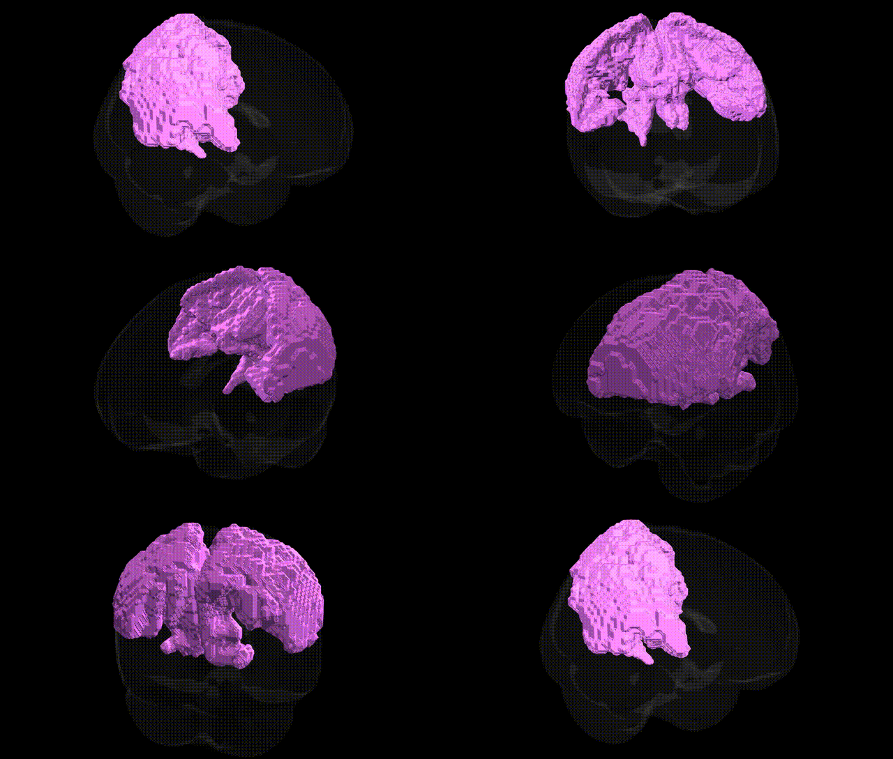
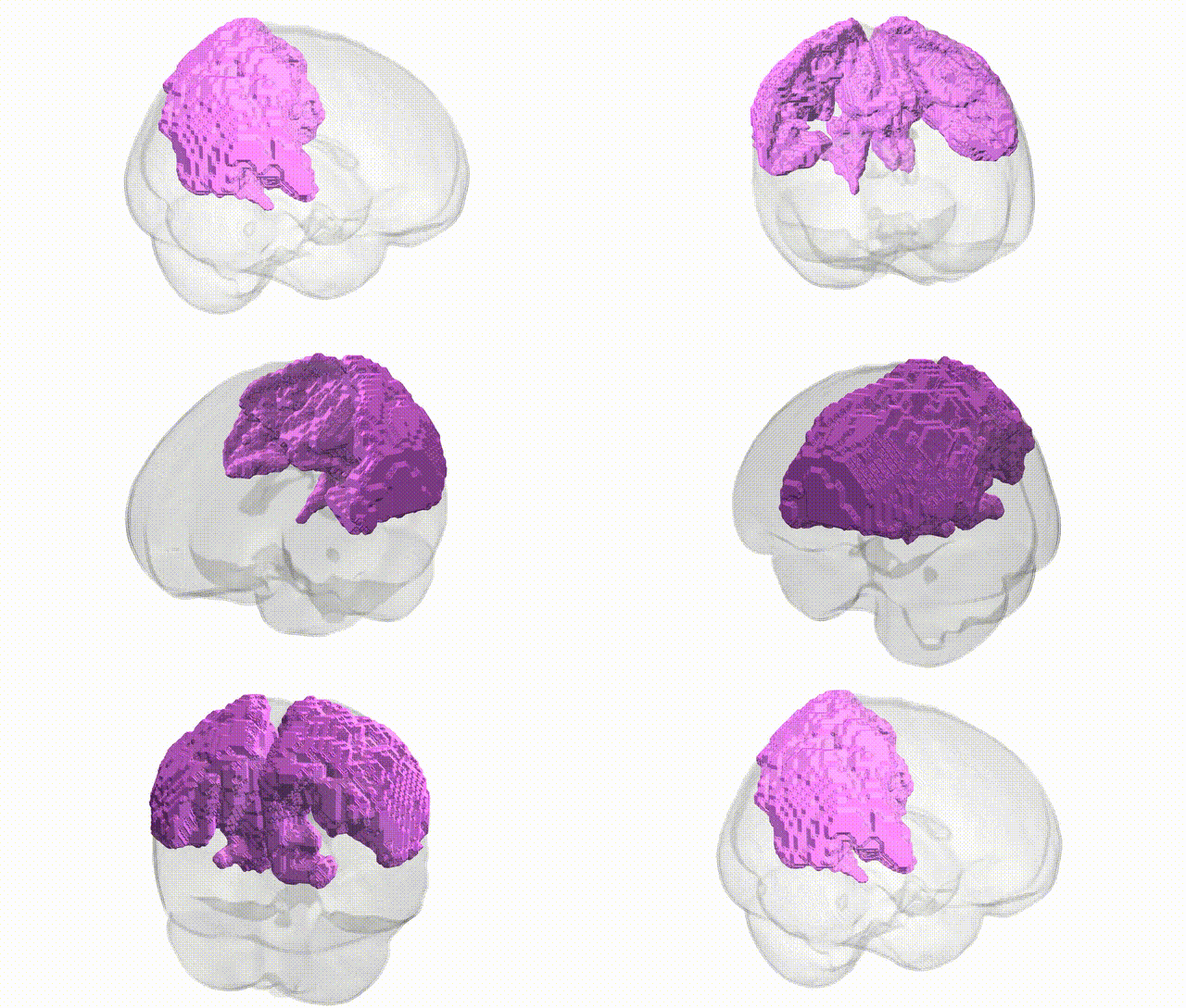
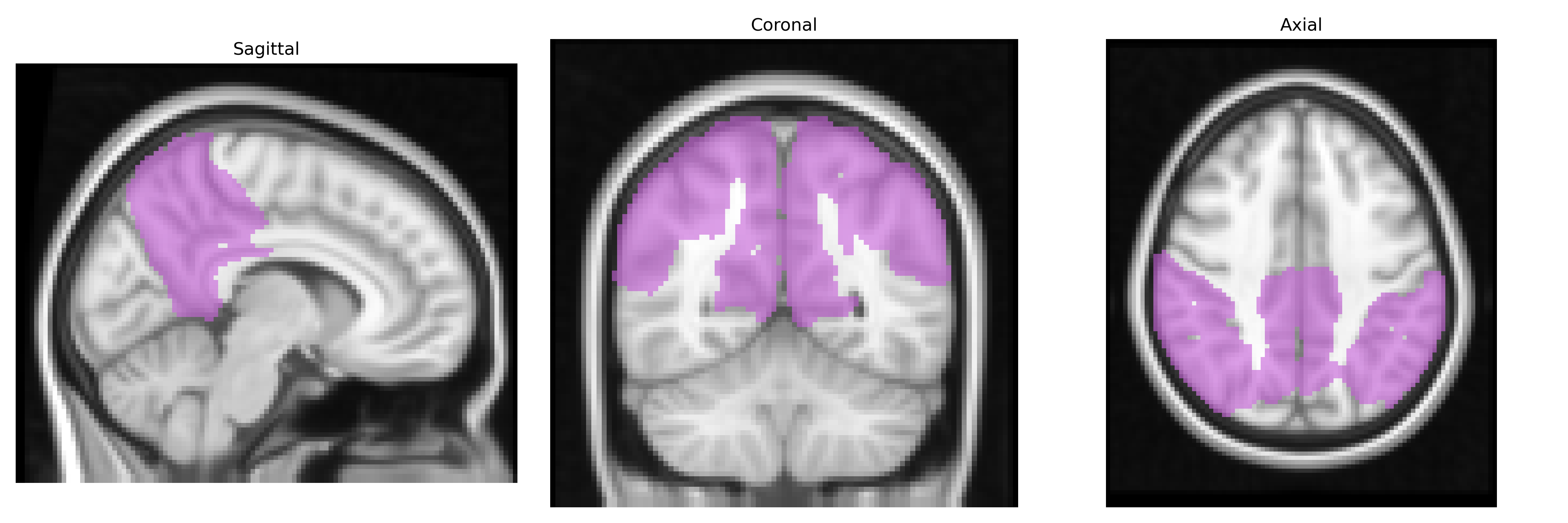
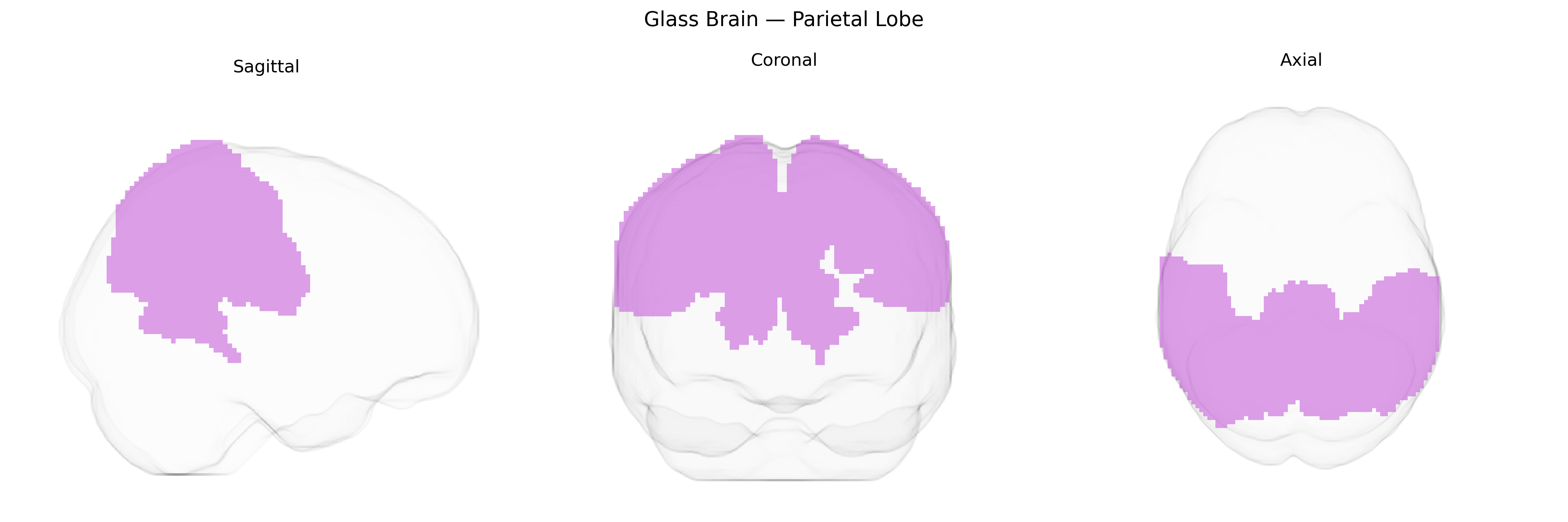
The bilateral parietal lobes comprise cortical regions located posterior to the central sulcus and superior to the occipital lobes in both hemispheres, encompassing the postcentral gyrus (primary somatosensory cortex), superior parietal lobule, and inferior parietal lobule. This region integrates multimodal sensory information, including tactile, proprioceptive, and visuospatial inputs, supporting functions such as body representation, spatial attention, sensorimotor coordination, and aspects of numerical and language processing. The parietal lobes contribute critically to perception of object location and movement, guidance of visually directed actions, and higher-order cognitive operations like mental rotation and spatial working memory. Damage to these areas can result in deficits such as hemispatial neglect, apraxia, impaired somatosensory discrimination, and disruptions in spatial cognition. Parietal lobe
The bilateral parietal lobes, as defined in the MNI Structural maxprob thr25 2 mm atlas (encompassing superior and inferior parietal regions including parts of the intraparietal sulcus and supramarginal/angular gyri), have been repeatedly implicated in genetic and GWAS-based studies of cognition, neurodevelopmental and psychiatric disorders, and brain structure. Polygenic scores for general cognitive ability and educational attainment, driven by loci such as those near genes including MAPT, KIBRA (WWC1), CHRNA4, and various synaptic and axon guidance genes, show associations with parietal cortical thickness, surface area, and functional activation in working memory, numerical processing, and visuospatial tasks. Large-scale imaging–genetics consortia (e.g., ENIGMA, UK Biobank) have identified SNP-based heritability and specific variants influencing parietal lobe morphometry and white matter integrity, involving genes related to neurodevelopment (e.g., NRG1, CNTNAP2, DISC1-like pathways) and neuronal signaling. GWAS of Alzheimer’s disease, frontotemporal dementia, and related endophenotypes link risk loci such as APOE, CLU, PICALM, and SORL1 with atrophy and hypometabolism in posterior parietal and precuneus regions, while schizophrenia and bipolar disorder risk variants (e.g., in MHC region, CACNA1C, ZNF804A) associate with altered parietal activation and connectivity. Autism spectrum disorder and ADHD genetic risk burden has been correlated with atypical parietal structure and network function, particularly in attention and default-mode–related parietal nodes. Additionally, GWAS of pain sensitivity, body image, and sensorimotor traits suggest parietal involvement via genes regulating somatosensory processing and cortical development, underscoring the parietal lobe as a genetically influenced hub for higher-order association, spatial cognition, and multimodal integration.
Overview generated by GPT-4o (2026).
Region ID: 6
Hemisphere: bilateral
Atlas: MNI Structural maxprob thr25 2mm
Full Quality Version: Download MP4
Full Quality Version: Download MP4
Wali Sidiqyar*, Gaurav Rudravaram*, Elyssa M. McMaster, Trent M. Schwartz, Adam M. Saunders, Kurt G. Schilling, Bennett A. Landman "Introducing SPINS: A Shared Public Visualization Library of Neuroanatomical Structures." Medical Imaging with Deep Learning- short paper
This resource is licensed under CC0 1.0 Universal (Public Domain).