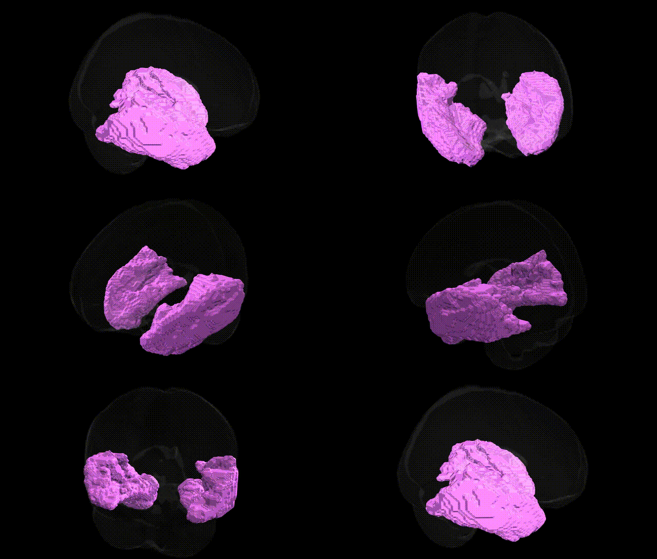
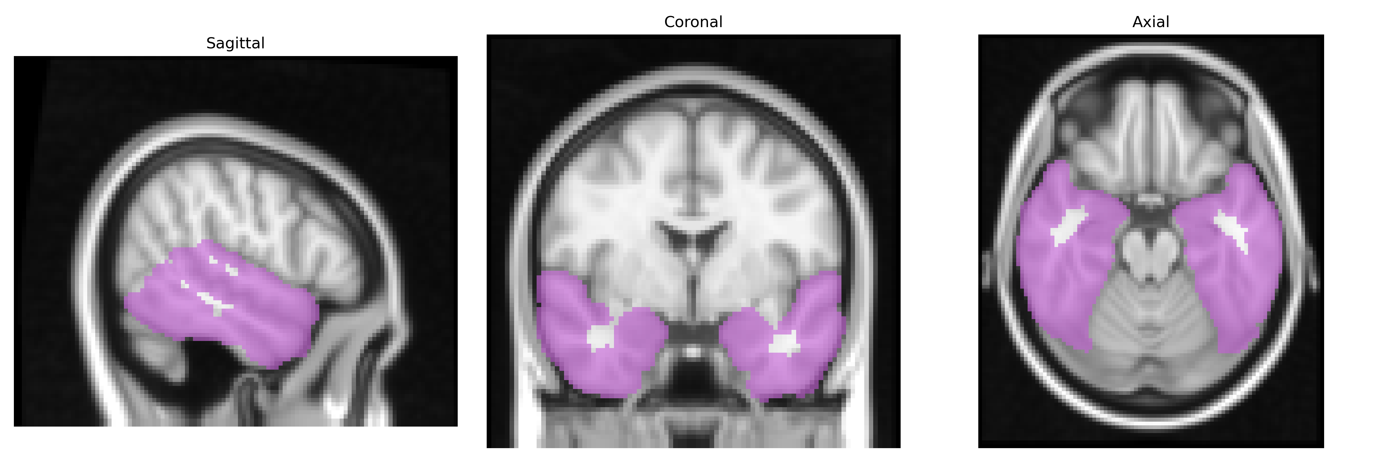
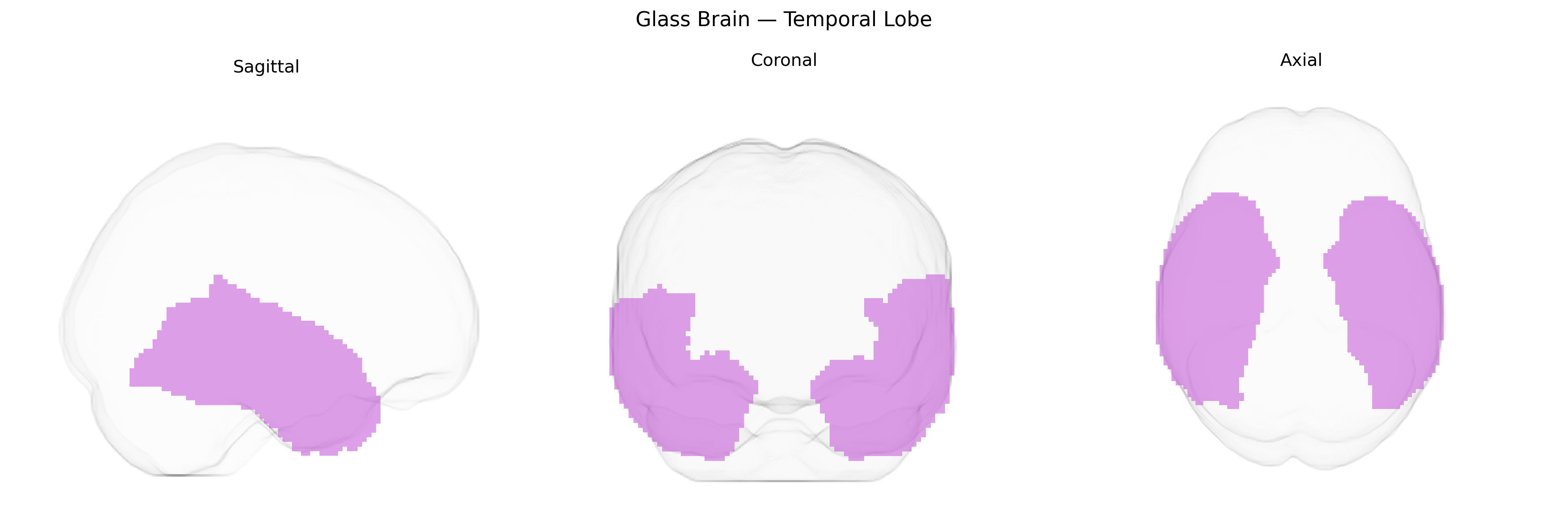
The bilateral temporal lobes are paired cortical regions located on the lateral aspects of the cerebral hemispheres, inferior to the lateral sulcus and anterior to the occipital lobes, and are primarily involved in auditory processing, language comprehension, semantic memory, and aspects of emotional and social cognition. Structurally, they encompass key gyri such as the superior, middle, and inferior temporal gyri, as well as deeper structures including portions of the limbic system, and are heavily interconnected with frontal and parietal association areas. Functionally, the temporal lobes are critical for the perception and interpretation of complex auditory stimuli, formation and retrieval of declarative memories, and integration of multimodal sensory information. Lesions in these regions can result in language deficits (e.g., Wernicke’s aphasia), impairments in memory and recognition, and changes in affect and social behavior. Temporal lobe
The bilateral temporal lobes, encompassing lateral temporal neocortex and medial temporal structures (including hippocampus and parahippocampal regions that overlap with the MNI Structural maxprob thr25 atlas), show robust genetic associations in large-scale GWAS and imaging genetics studies, particularly for cortical thickness and surface area, hippocampal volume, and temporal pole morphology, with key loci including variants in or near genes such as MAPT, KIAA0586, WNT3, DLG2, MSRB3, and multiple 17q21.31 haplotype-region genes. Heritability estimates for temporal lobe measures are high (often >40–60%), and specific SNPs have been repeatedly linked to individual differences in temporal lobe structure and connectivity in UK Biobank and ENIGMA consortia analyses. Pathology-related genetic studies implicate temporal lobe structures in Alzheimer’s disease (e.g., APOE, BIN1, CLU, CR1, TREM2 variants associated with atrophy in medial temporal regions and episodic memory decline), temporal lobe epilepsy (genes including LGI1, RELN, GABRG2, and others influencing seizure susceptibility and hippocampal sclerosis), frontotemporal dementia and semantic variant primary progressive aphasia (MAPT and GRN, with selective anterior temporal lobe degeneration), and schizophrenia and bipolar disorder (risk loci in CACNA1C, ZNF804A, and regions on 3p, 10q, and 22q associated with temporal lobe volume and functional abnormalities). Additional GWAS link temporal lobe structure and function to language and reading abilities, general cognitive performance, and auditory processing traits, indicating that polygenic influences on these behaviors are partly mediated through variation in temporal lobe anatomy and connectivity.
Overview generated by GPT-4o (2026).
Region ID: 8
Hemisphere: bilateral
Atlas: MNI Structural maxprob thr25 2mm
Full Quality Version: Download MP4
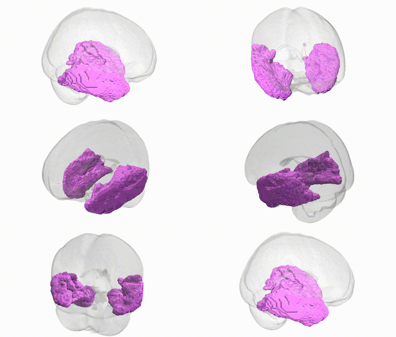
Full Quality Version: Download MP4
Wali Sidiqyar*, Gaurav Rudravaram*, Elyssa M. McMaster, Trent M. Schwartz, Adam M. Saunders, Kurt G. Schilling, Bennett A. Landman "Introducing SPINS: A Shared Public Visualization Library of Neuroanatomical Structures." Medical Imaging with Deep Learning- short paper
This resource is licensed under CC0 1.0 Universal (Public Domain).