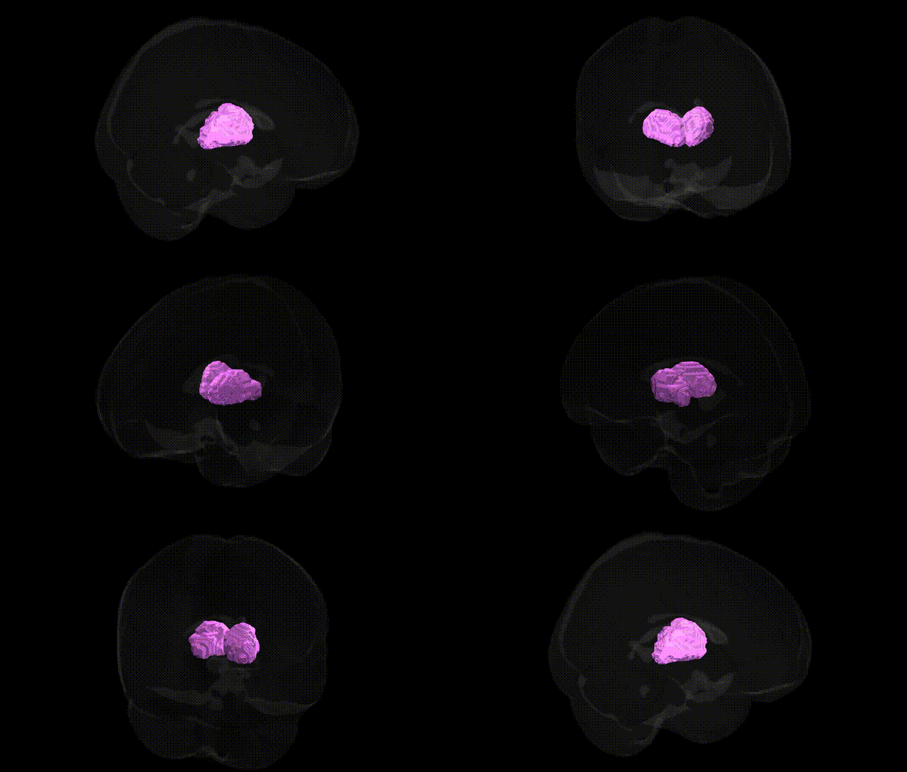
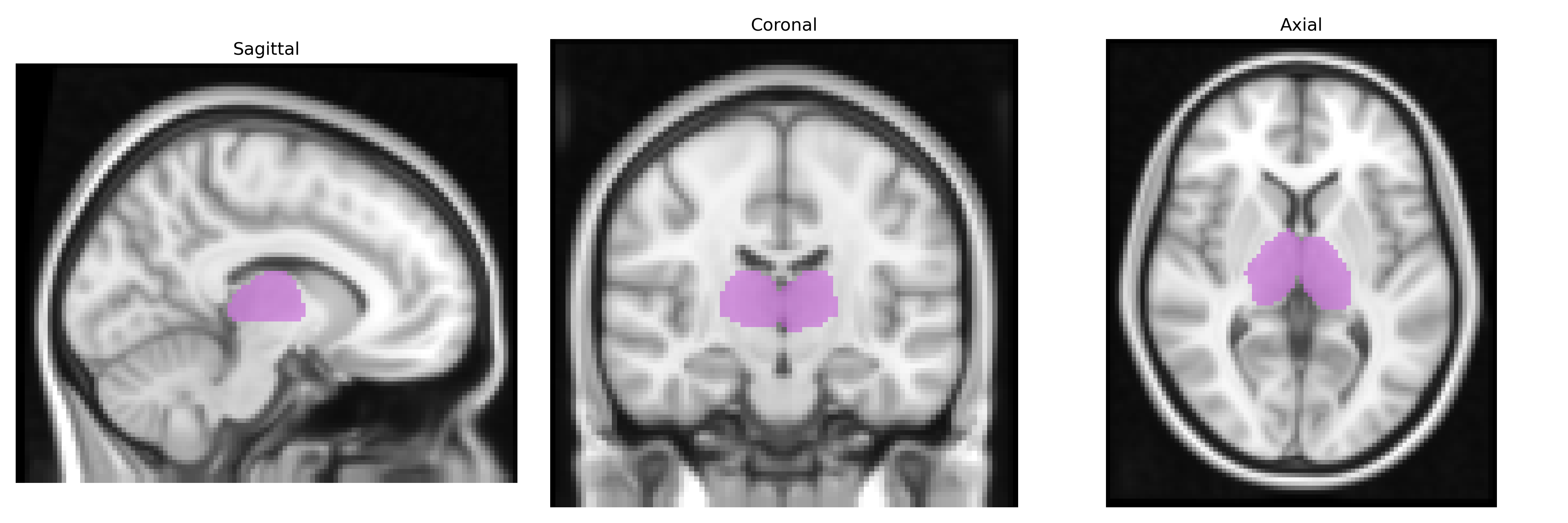
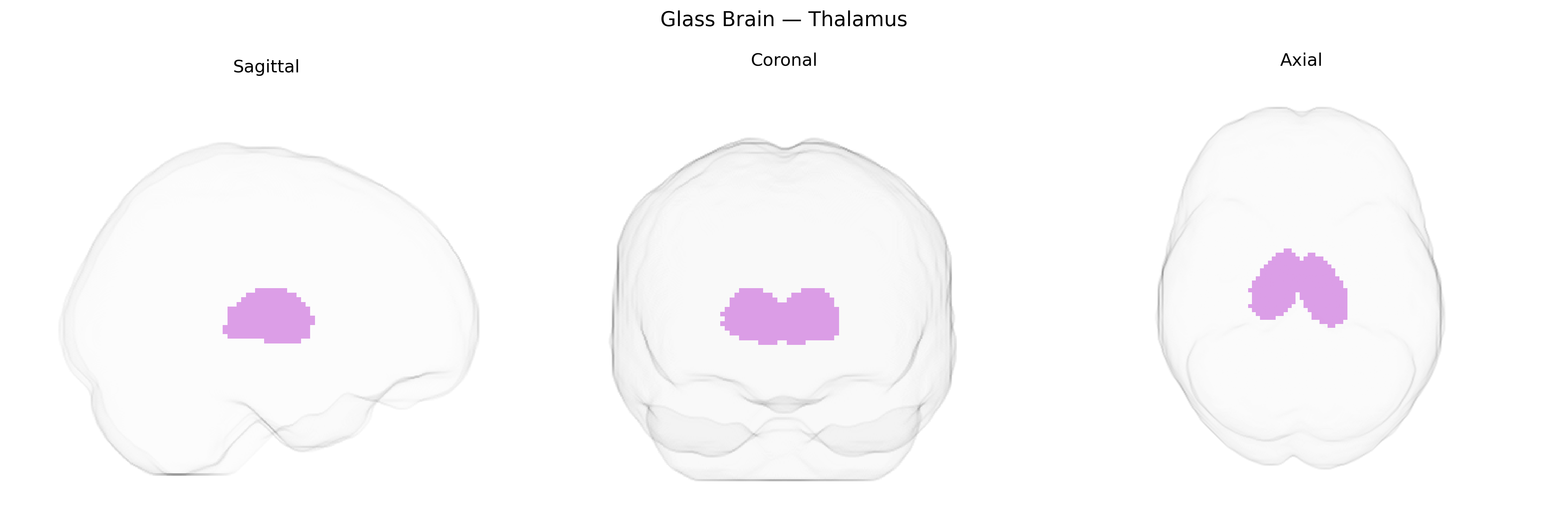
The bilateral thalamus is a paired, ovoid diencephalic structure situated on either side of the third ventricle, serving as the principal relay hub for sensory, motor, and integrative information between subcortical regions and the cerebral cortex. Composed of multiple nuclei (including relay, association, and intralaminar nuclei), it processes and modulates inputs from sensory pathways (except olfaction), basal ganglia, cerebellum, and limbic structures, contributing critically to perception, motor control, arousal, attention, and consciousness. Functionally, the thalamus organizes and gates information flow, refining signal transmission through reciprocal corticothalamic connections and neuromodulatory influences. Within the MNI Structural maxprob thr25 2mm Atlas, the bilateral thalamus region represents voxels most likely belonging to thalamic tissue in both hemispheres, capturing its core anatomical boundaries and ensuring consistent localization for structural and functional neuroimaging analyses. Thalamus
Genetic studies implicating the bilateral thalamus—including regions defined in the MNI Structural maxprob thr25 2mm Atlas—have identified multiple loci influencing thalamic volume, connectivity, and function, with several robust findings from large-scale GWAS of subcortical brain structures. Common variants near genes such as CRTC1, DCC, MAPT, and others involved in neurodevelopment, synaptic signaling, and axonal guidance have been associated with thalamic size and microstructure, often overlapping with loci that influence global brain morphology and cognitive phenotypes. Polygenic signals linked to schizophrenia, bipolar disorder, major depressive disorder, and autism spectrum disorder show enrichment in genes affecting thalamocortical circuitry, consistent with imaging genetics findings that thalamic abnormalities are central to these conditions. Thalamic volume and integrity have also been genetically correlated with traits such as general cognitive ability, educational attainment, sleep regulation, and risk for neurodegenerative diseases like Alzheimer’s and Parkinson’s disease, with risk variants in APOE, MAPT, and other pathways linked to thalamic atrophy or altered connectivity. Overall, GWAS and related imaging-genetic approaches support a highly polygenic architecture in which neurodevelopmental, synaptic, and immune-related genes jointly modulate thalamic structure and function, contributing to vulnerability for a broad spectrum of psychiatric, cognitive, and neurological traits.
Overview generated by GPT-4o (2026).
Region ID: 9
Hemisphere: bilateral
Atlas: MNI Structural maxprob thr25 2mm
Full Quality Version: Download MP4
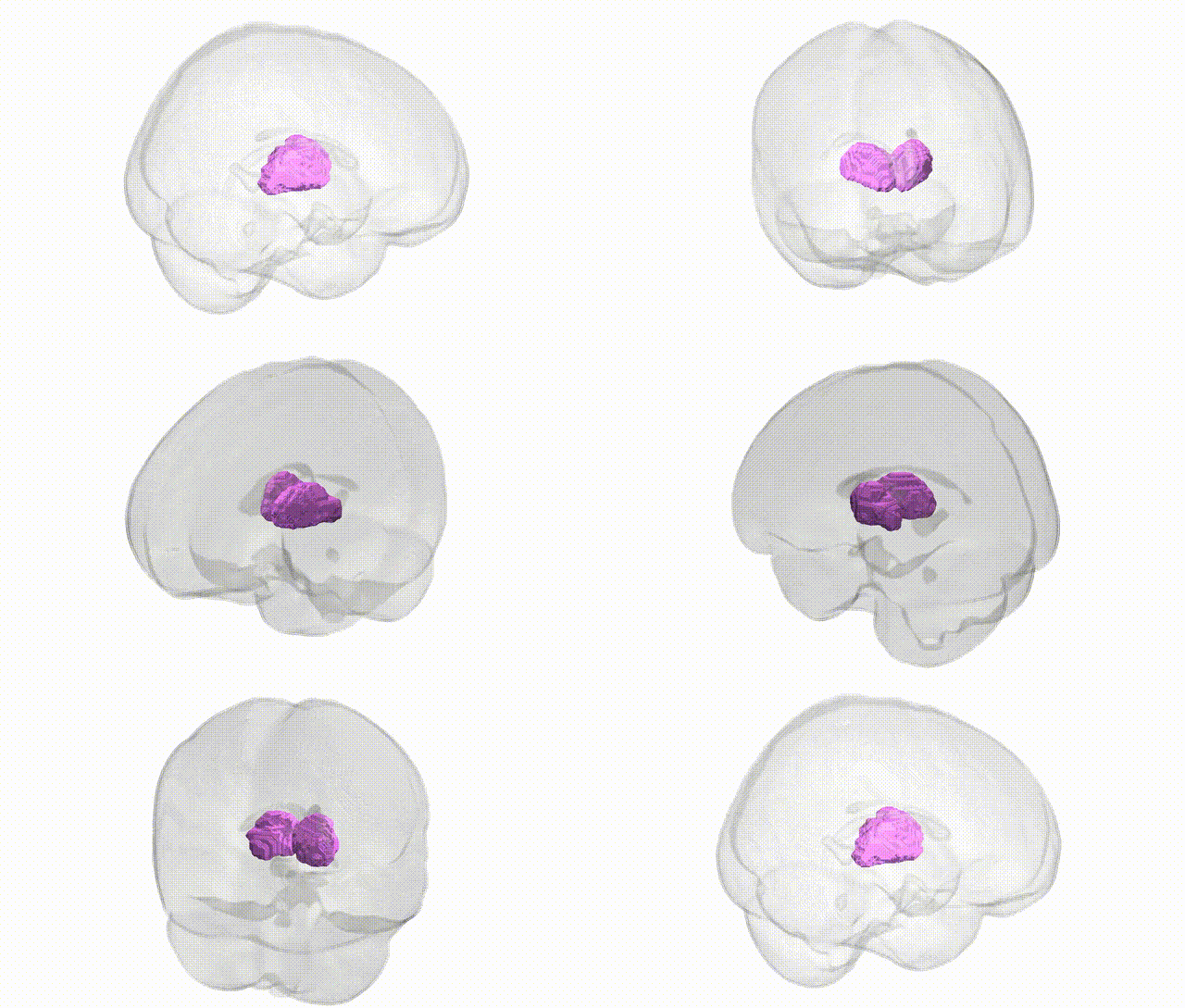
Full Quality Version: Download MP4
Wali Sidiqyar*, Gaurav Rudravaram*, Elyssa M. McMaster, Trent M. Schwartz, Adam M. Saunders, Kurt G. Schilling, Bennett A. Landman "Introducing SPINS: A Shared Public Visualization Library of Neuroanatomical Structures." Medical Imaging with Deep Learning- short paper
This resource is licensed under CC0 1.0 Universal (Public Domain).