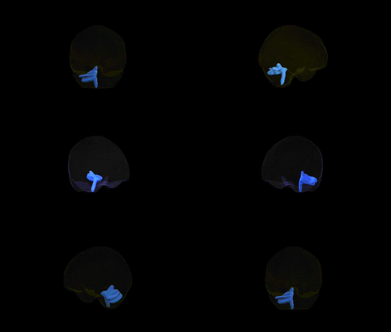
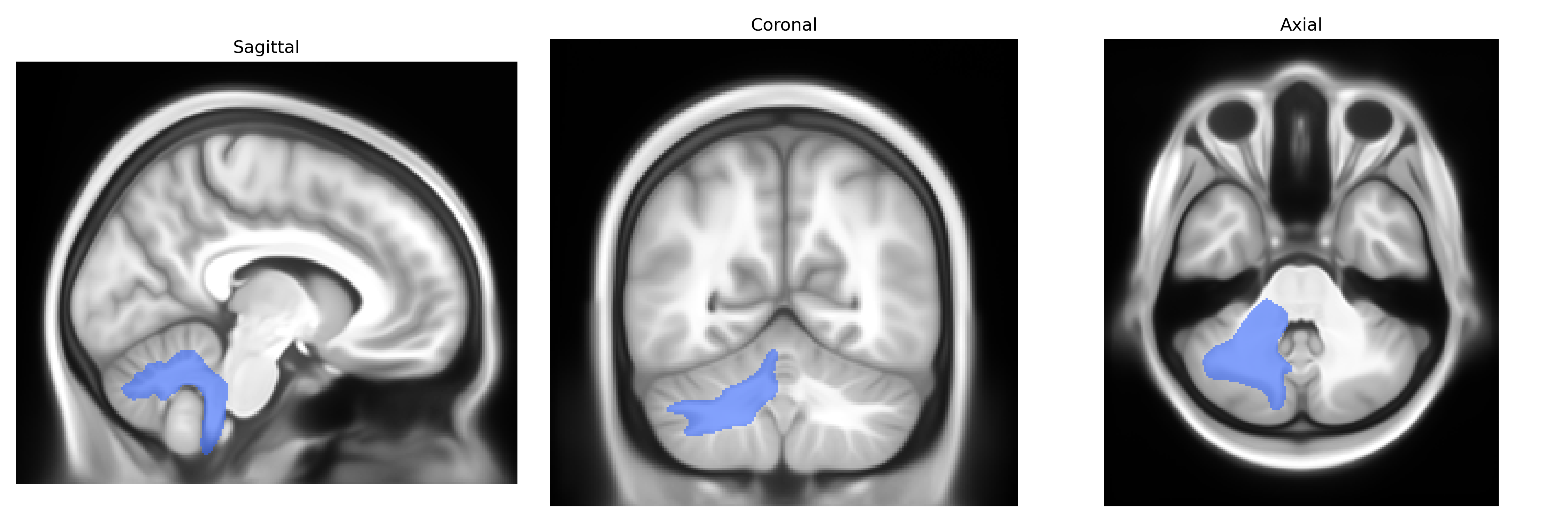
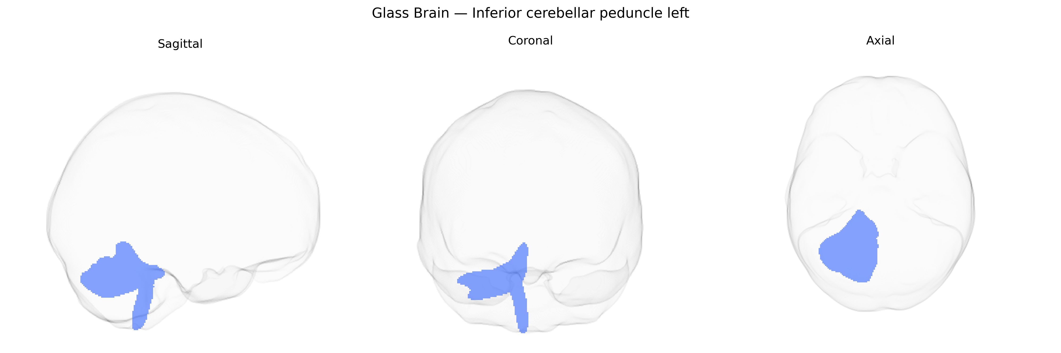
The Inferior cerebellar peduncle left is a major white matter tract connecting the medulla oblongata and spinal cord to the ipsilateral cerebellar hemisphere, primarily conveying afferent fibers involved in proprioception, balance, and vestibular functions. It carries inputs from the dorsal spinocerebellar and cuneocerebellar tracts, the inferior olivary nucleus (olivocerebellar fibers), the vestibular nuclei, and reticular formation, projecting mainly to the cerebellar cortex and deep cerebellar nuclei, particularly influencing coordination of limb movements and postural control. This tract plays a critical role in integrating sensory feedback with motor commands to refine ongoing movements and maintain equilibrium; lesions typically result in ipsilateral ataxia, dysmetria, and gait disturbances. There is no direct link for the unilateral tract; a related structure is the Inferior cerebellar peduncle.
Current literature provides very limited tract-specific genetic information for the left inferior cerebellar peduncle (ICP) as defined in the Pandora‑TractSeg Atlas; most diffusion MRI GWAS aggregate cerebellar or brainstem tracts rather than isolating this structure, and few, if any, large-scale studies report genome-wide significant loci explicitly annotated to the left ICP. Broad diffusion tensor imaging GWAS have identified polygenic influences on fractional anisotropy and mean diffusivity in cerebellar and brainstem white matter, often implicating genes involved in axonal guidance, myelination, and neurodevelopment (for example, loci near genes such as NTRK3, CNTN4, or PLP1 in related tracts), but these associations are generally reported at the level of composite cerebellar or inferior cerebellar peduncle regions rather than the left tract alone. Clinical and imaging studies link inferior cerebellar peduncle microstructure to ataxias, developmental coordination disorder, autism spectrum disorder, and schizophrenia, yet these findings are usually phenotypic (differences in FA/MD or volume by diagnosis or polygenic risk score) and do not provide tract‑specific GWAS hits or well-replicated gene–tract associations restricted to the left ICP. Overall, as of current knowledge, robust, reproducible genetic associations uniquely and specifically mapped to the left inferior cerebellar peduncle in the Pandora‑TractSeg Atlas remain largely uncharacterized, and most insights are extrapolated from broader cerebellar or cerebellar‑peduncle white matter analyses.
Overview generated by GPT-4o (2026).
Region ID: 21
Hemisphere: left
Atlas: Pandora-TractSeg
Full Quality Version: Download MP4
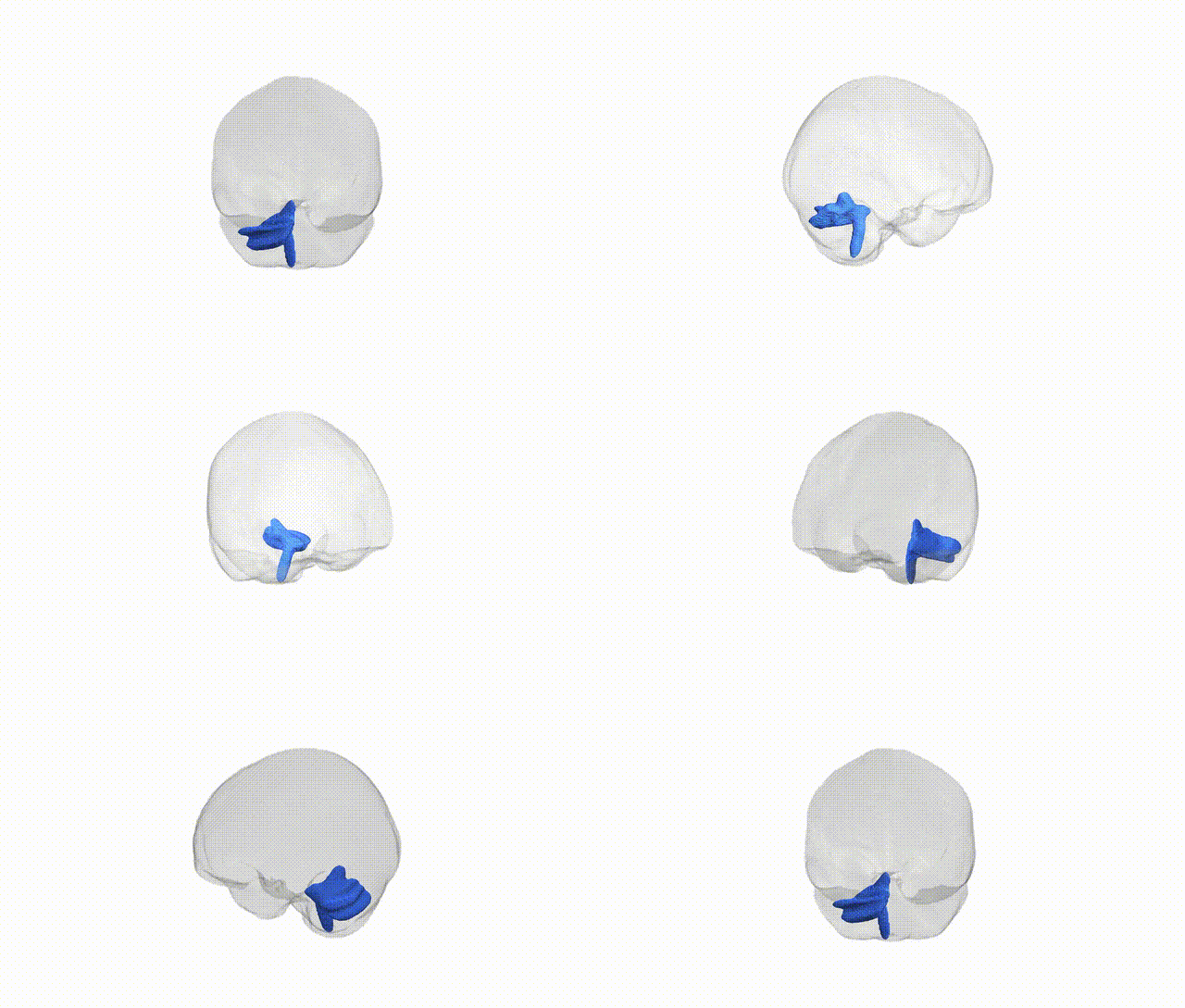
Full Quality Version: Download MP4
Wali Sidiqyar*, Gaurav Rudravaram*, Elyssa M. McMaster, Trent M. Schwartz, Adam M. Saunders, Kurt G. Schilling, Bennett A. Landman "Introducing SPINS: A Shared Public Visualization Library of Neuroanatomical Structures." Medical Imaging with Deep Learning- short paper
This resource is licensed under CC0 1.0 Universal (Public Domain).