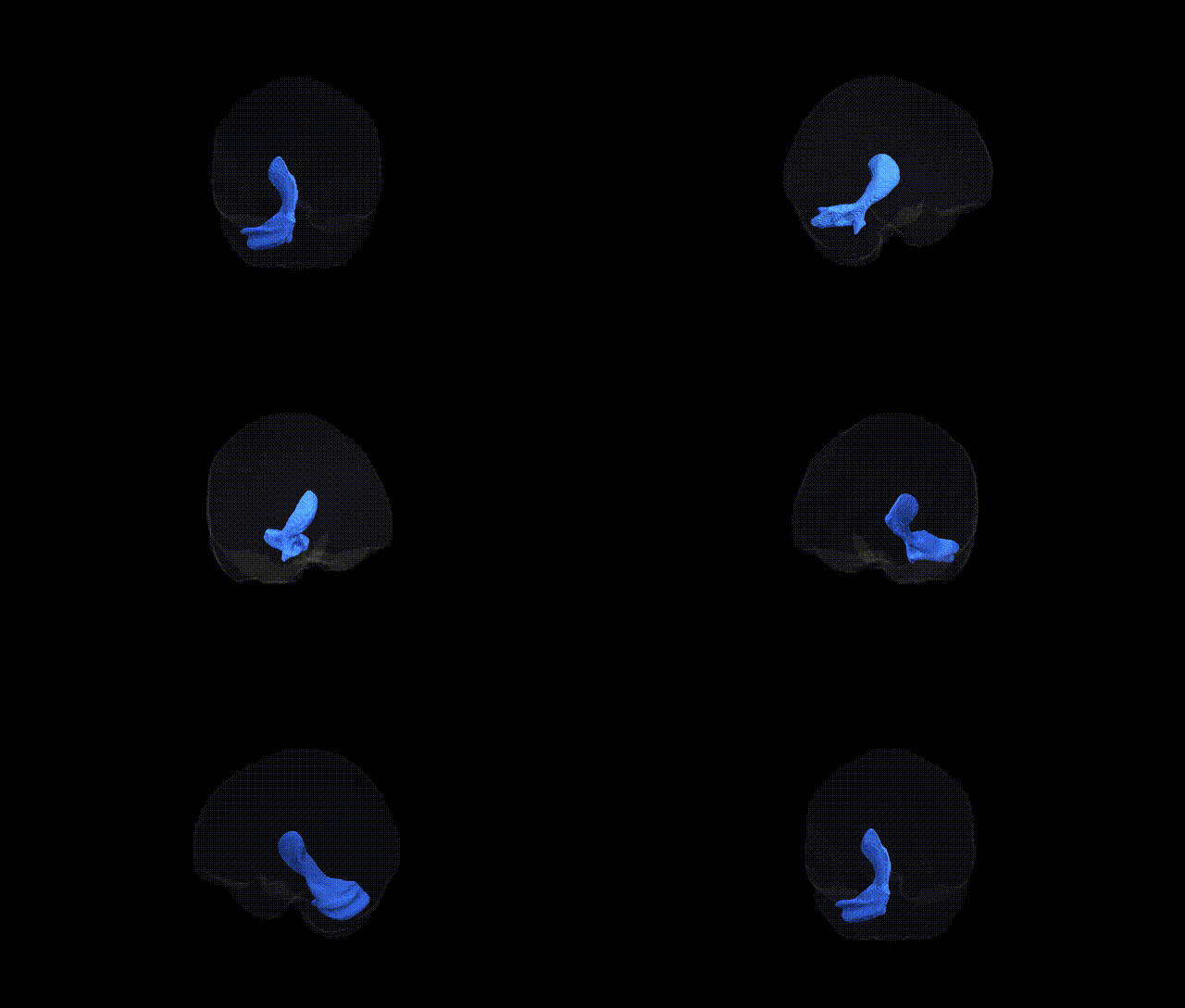
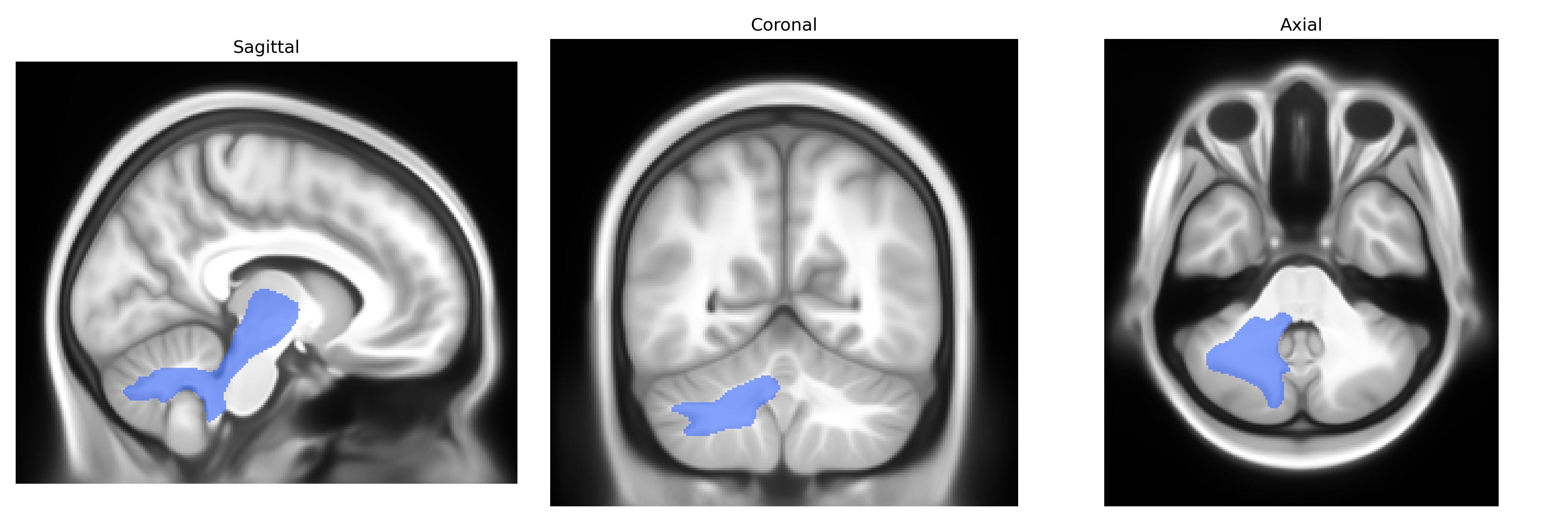
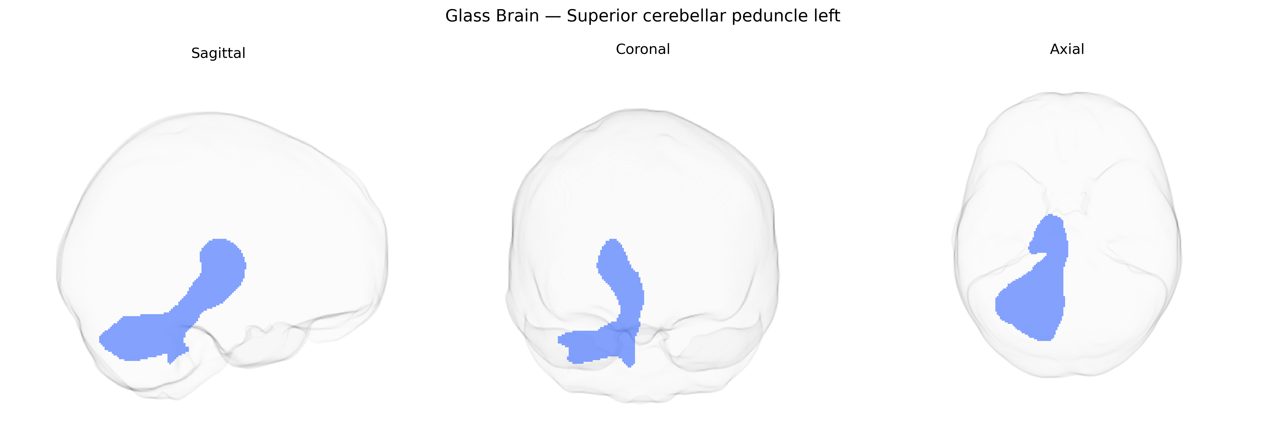
The Superior cerebellar peduncle left is a major efferent white matter tract connecting the cerebellum to the midbrain, forming the principal pathway by which cerebellar output reaches premotor and motor structures. Arising predominantly from neurons in the dentate, interposed, and fastigial nuclei of the left cerebellar hemisphere, its fibers ascend rostrally, decussate in the midbrain at the level of the inferior colliculi, and project mainly to the red nucleus and ventrolateral thalamic nuclei, which in turn relay signals to widespread cortical motor and premotor areas. This tract plays a key role in the coordination, planning, and fine-tuning of voluntary movement, motor learning, and aspects of motor timing, and its disruption can lead to ataxia, dysmetria, and other cerebellar motor syndromes. No direct link exists for the left tract specifically; see the related structure Superior cerebellar peduncle.
Current genetic knowledge specifically targeting the left superior cerebellar peduncle white matter tract as defined in the Pandora‑TractSeg Atlas is very limited, and no robust GWAS has yet isolated variants uniquely associated with this tract independent of nearby or global cerebellar and brainstem pathways. Large diffusion MRI GWAS (e.g., UK Biobank–based studies) have identified numerous loci influencing cerebellar peduncle diffusion measures such as fractional anisotropy and mean diffusivity, but typically aggregate across bilateral or combined superior cerebellar peduncles and often do not provide tract-specific left-sided results or direct mapping to the Pandora‑TractSeg framework. Reported loci often involve genes with broad roles in neurodevelopment, axonal guidance, and myelination (for example, variants near or within genes such as MAG, NRXN1, or others implicated in white matter microstructure), and these cerebellar peduncle measures have been linked in polygenic and phenotypic analyses to cognitive performance, motor coordination, and risk for neuropsychiatric or neurodevelopmental conditions (including schizophrenia, ADHD, and autism spectrum disorder), but these links are generally nonspecific to the left superior cerebellar peduncle. Overall, existing evidence supports a heritable and genetically influenced component to microstructural variation in the superior cerebellar peduncles, yet precise, tract- and hemisphere-specific genetic associations for the left superior cerebellar peduncle within the Pandora‑TractSeg Atlas remain largely uncharacterized in the current literature.
Overview generated by GPT-4o (2026).
Region ID: 34
Hemisphere: left
Atlas: Pandora-TractSeg
Full Quality Version: Download MP4
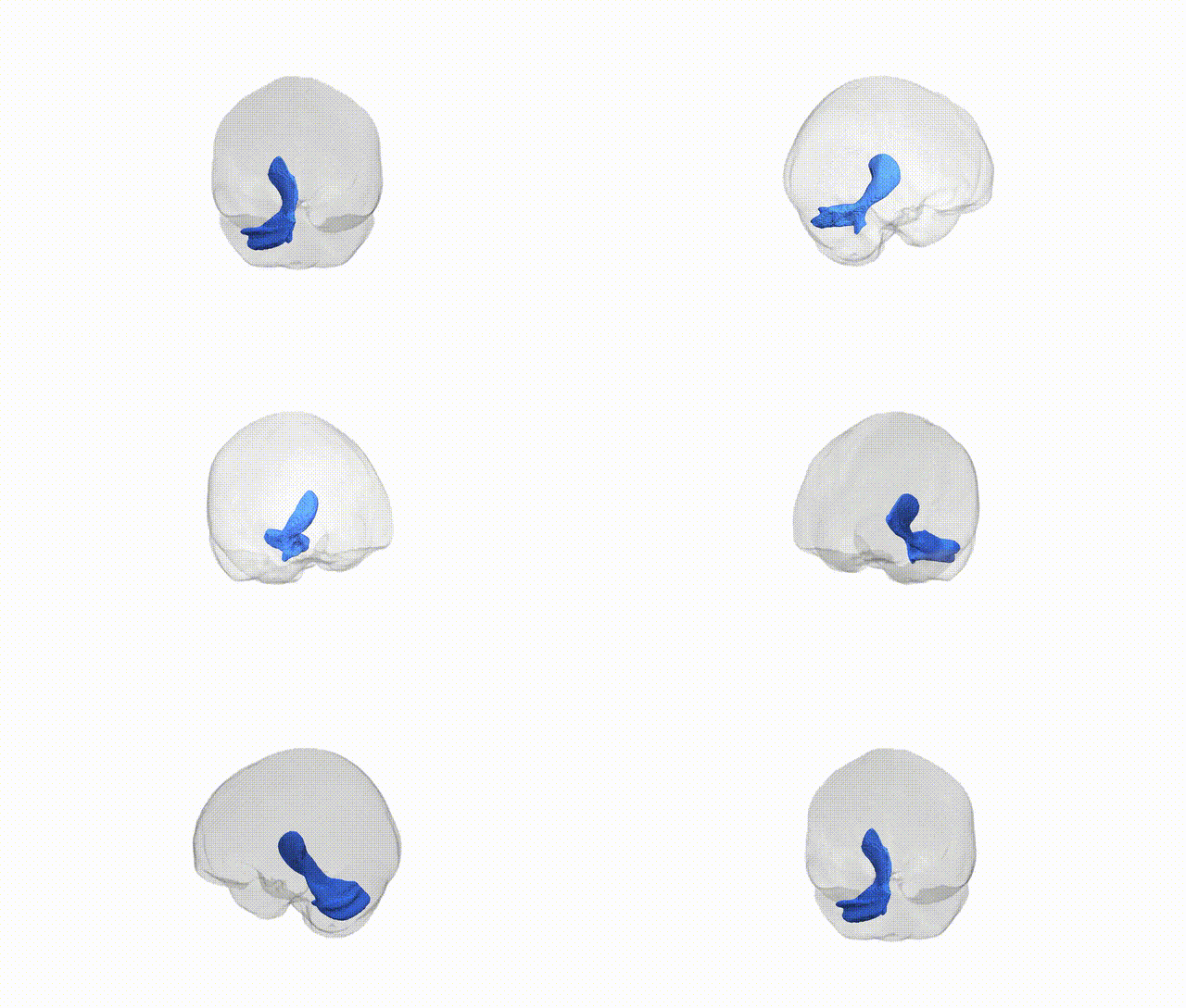
Full Quality Version: Download MP4
Wali Sidiqyar*, Gaurav Rudravaram*, Elyssa M. McMaster, Trent M. Schwartz, Adam M. Saunders, Kurt G. Schilling, Bennett A. Landman "Introducing SPINS: A Shared Public Visualization Library of Neuroanatomical Structures." Medical Imaging with Deep Learning- short paper
This resource is licensed under CC0 1.0 Universal (Public Domain).