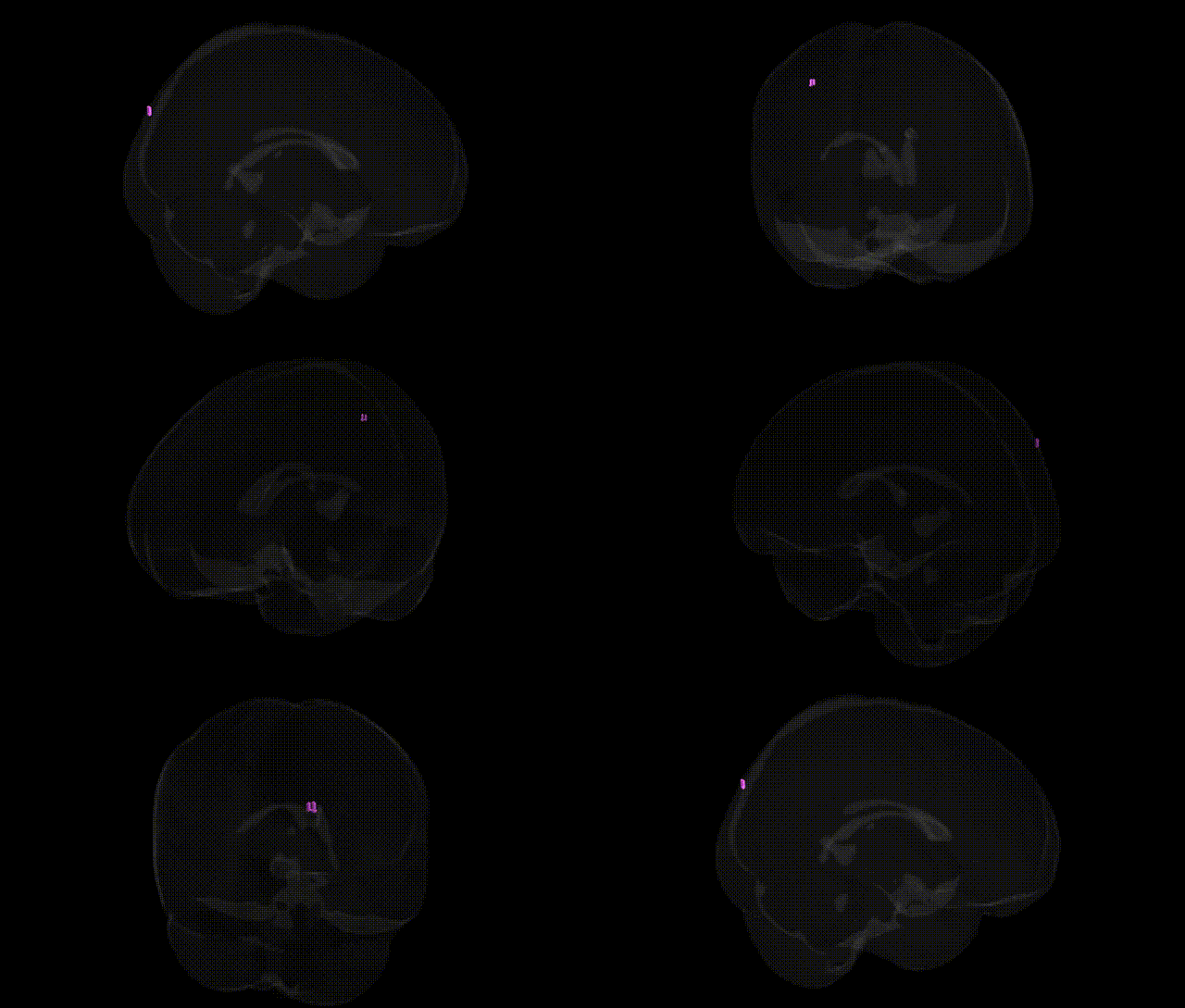
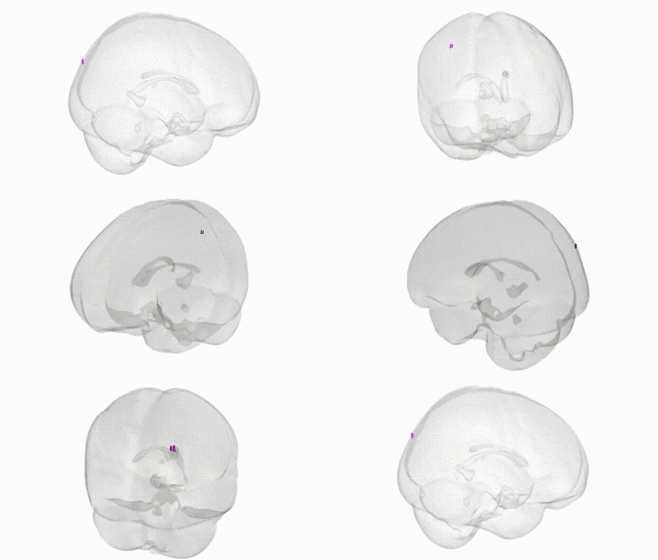
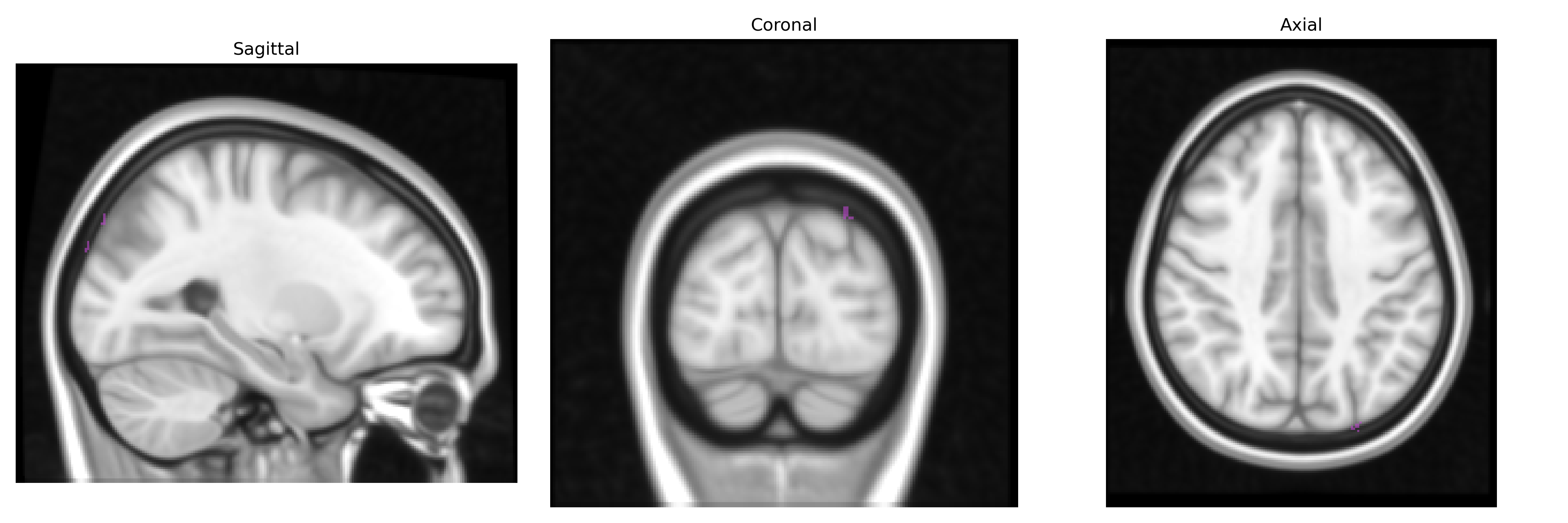
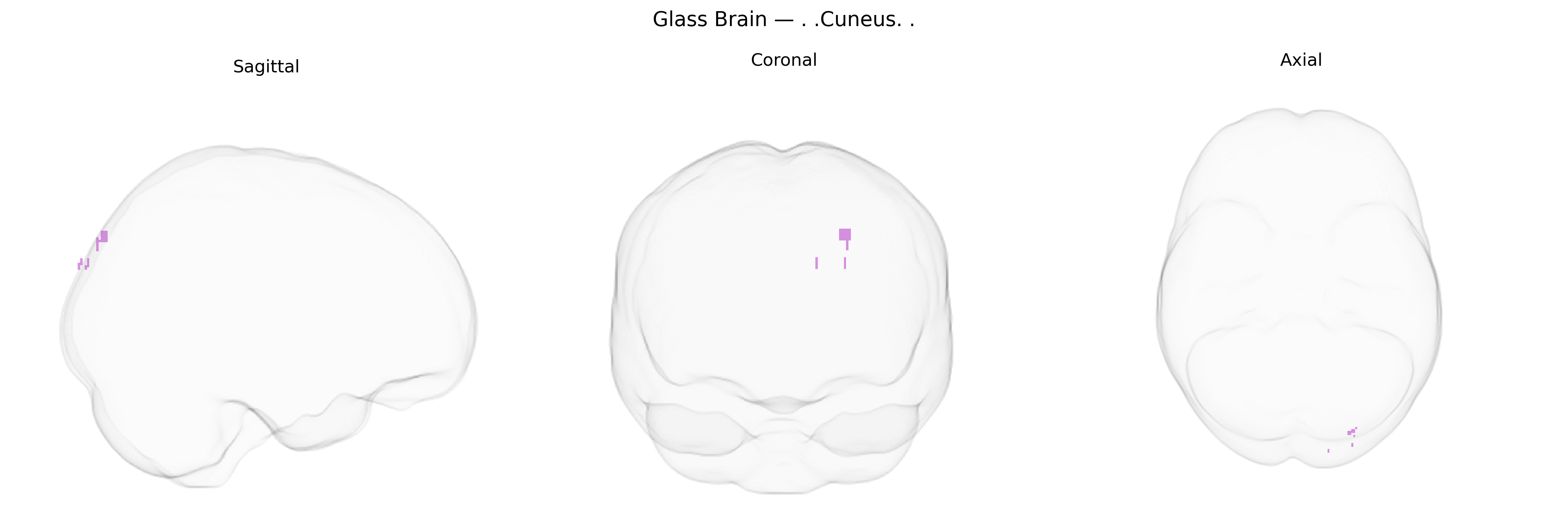
The bilateral cuneus is a wedge-shaped region of the occipital lobe located on the medial surface of the cerebral hemisphere, bounded superiorly by the parieto-occipital sulcus and inferiorly by the calcarine sulcus. It is primarily involved in early-stage visual processing, including basic features such as orientation, spatial frequency, and motion, and contributes to visuospatial integration and visual attention. The cuneus receives major input from the primary visual cortex and higher-order visual association areas, participating in the dorsal visual stream that supports spatial awareness and visually guided behavior. Lesions affecting the cuneus can lead to defects in the corresponding portions of the visual field, reflecting its retinotopic organization. Cuneus
The bilateral cuneus, a medial occipital lobe region crucial for basic visual processing and visuospatial integration, has been implicated in several genetic imaging and GWAS findings, though typically as part of broader occipital or visual cortex measures rather than as an isolated Talairach 1 mm Atlas label. Variants near genes involved in synaptic plasticity and neurodevelopment—such as BDNF, GRIN2B, and NRGN—have been associated with cuneus gray matter volume or cortical thickness in neuroimaging genetics studies, often in the context of visual processing efficiency and general cognitive function. Large-scale GWAS of brain imaging phenotypes (e.g., ENIGMA, UK Biobank) have identified multiple loci influencing occipital cortical structure and functional activation, with cuneus metrics contributing to polygenic architecture of traits like intelligence, educational attainment, and visual attention. In psychiatric and neurological disorders, cuneus structural and functional alterations have been reported in schizophrenia, major depressive disorder, bipolar disorder, autism spectrum disorder, and Alzheimer’s disease, with some disorder-associated risk variants (for example within CACNA1C, ZNF804A, APOE) showing downstream effects on occipital/cuneus activation or morphology in patient and high-risk cohorts. Additionally, GWAS of visual-related traits (e.g., myopia, migraine with aura, and visual hallucinations in Parkinson’s disease or Lewy body dementia) have occasionally linked risk alleles to altered activity or connectivity in cuneus-inclusive visual networks, suggesting that genetic variation in neurodevelopmental, synaptic, and vascular pathways can modulate structure and function of this region and thereby contribute to disease vulnerability and cognitive/visual phenotypes.
Overview generated by GPT-4o (2026).
Region ID: 842
Hemisphere: bilateral
Atlas: Talairach labels 1mm
Full Quality Version: Download MP4
Full Quality Version: Download MP4
Wali Sidiqyar*, Gaurav Rudravaram*, Elyssa M. McMaster, Trent M. Schwartz, Adam M. Saunders, Kurt G. Schilling, Bennett A. Landman "Introducing SPINS: A Shared Public Visualization Library of Neuroanatomical Structures." Medical Imaging with Deep Learning- short paper
This resource is licensed under CC0 1.0 Universal (Public Domain).