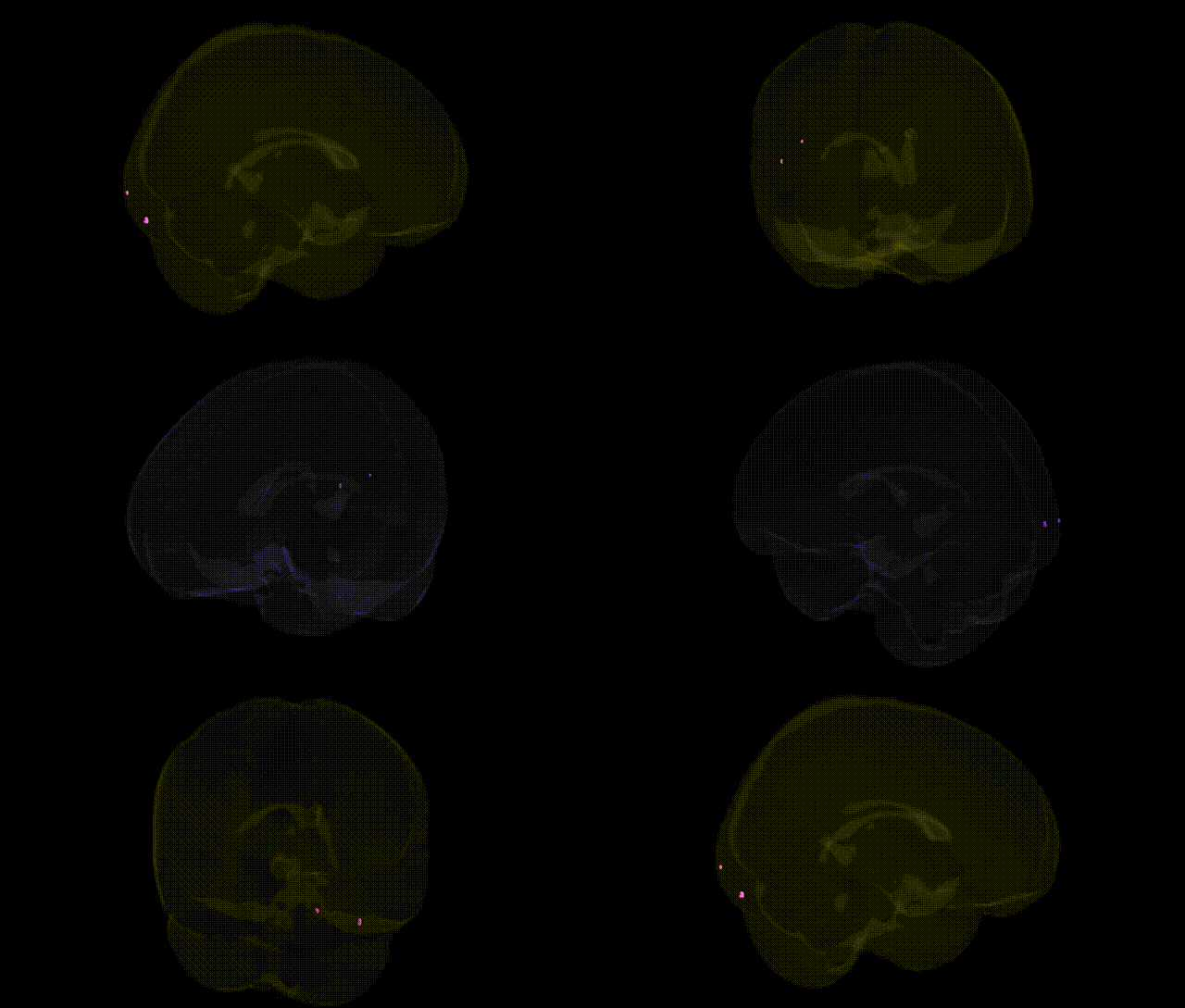
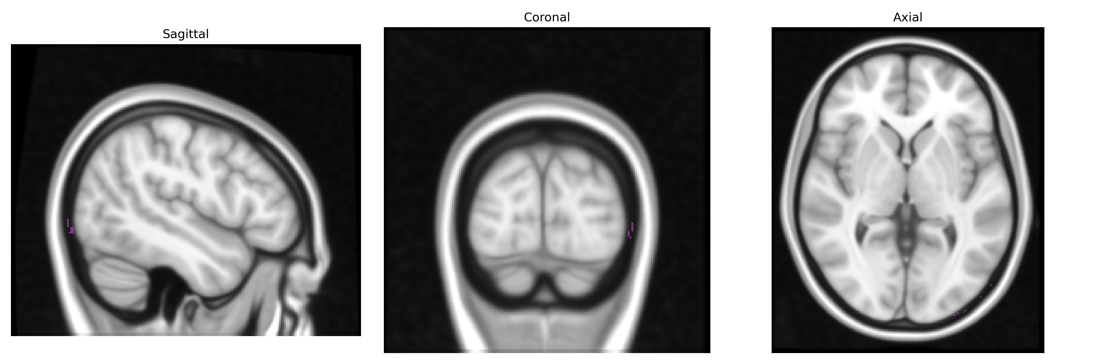
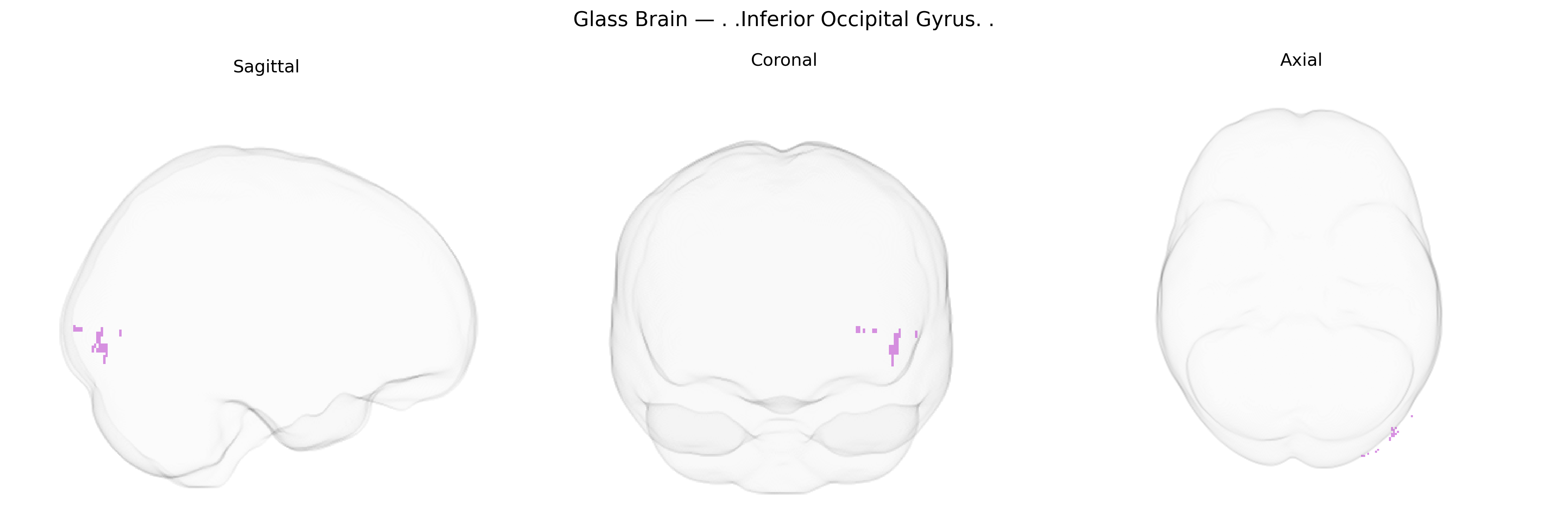
The bilateral Inferior Occipital Gyrus is a cortical region located in the ventral portion of the occipital lobe, bordering the temporal lobe and forming part of the visual association cortex involved in early and intermediate stages of visual processing. It lies inferior to the middle occipital gyrus and contributes to the analysis of complex visual features, including shape, contour, and object-related information, with inputs from primary visual areas (V1) and projections toward ventral visual stream structures. Functionally, the Inferior Occipital Gyrus participates in the integration of visual stimuli that support object recognition and may be involved in processing faces and other category-specific visual information as part of broader occipitotemporal networks. There is no direct Wikipedia article for the Inferior Occipital Gyrus; a closely related and encompassing structure is the occipital lobe: Occipital lobe.
The bilateral Inferior Occipital Gyrus (IOG), a key visual association area, appears in imaging-genetics studies primarily through genome-wide associations with occipital cortical thickness, surface area, and functional activation rather than via Talairach-specific labels per se. Large GWAS of brain morphology (e.g., ENIGMA and UK Biobank) have identified multiple loci—often near genes involved in neurodevelopment, synaptic function, and axon guidance (such as variants around HMGA2, FOXO3, and pathways including Wnt and MAPK signaling)—that influence occipital lobe structure encompassing regions overlapping the IOG. Occipital measures that include the IOG have been genetically correlated with general cognitive ability, educational attainment, and visual processing traits, as well as with neuropsychiatric conditions like schizophrenia, major depression, and autism spectrum disorder, suggesting pleiotropic effects of shared variants on visual cortex development and higher-order cognition. Imaging-genetics work in dyslexia and reading ability has reported associations between risk variants (e.g., in DCDC2, KIAA0319, and other neuronal migration genes) and functional or structural differences in occipito-temporal areas that often include the inferior occipital region as part of the visual word form network. Additionally, GWAS of face recognition and social cognition have implicated occipito-temporal activation patterns influenced by polygenic scores, indirectly linking genetic risk for social and affective disorders to variation in IOG recruitment during visual tasks. Overall, genetic associations involving the Inferior Occipital Gyrus emerge mainly through broader occipital or occipito-temporal phenotypes, with convergent evidence that common variants affecting neurodevelopment and synaptic plasticity modulate the structure and function of this region and thereby contribute to individual differences in visual cognition and vulnerability to psychiatric and neurodevelopmental disorders.
Overview generated by GPT-4o (2026).
Region ID: 260
Hemisphere: bilateral
Atlas: Talairach labels 1mm
Full Quality Version: Download MP4
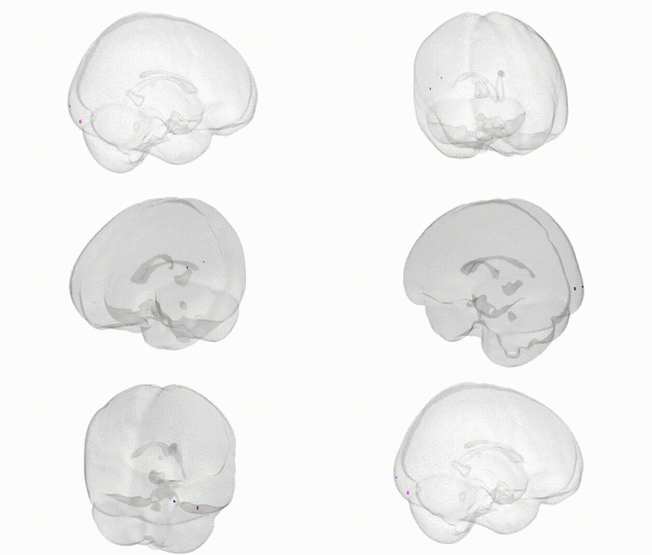
Full Quality Version: Download MP4
Wali Sidiqyar*, Gaurav Rudravaram*, Elyssa M. McMaster, Trent M. Schwartz, Adam M. Saunders, Kurt G. Schilling, Bennett A. Landman "Introducing SPINS: A Shared Public Visualization Library of Neuroanatomical Structures." Medical Imaging with Deep Learning- short paper
This resource is licensed under CC0 1.0 Universal (Public Domain).