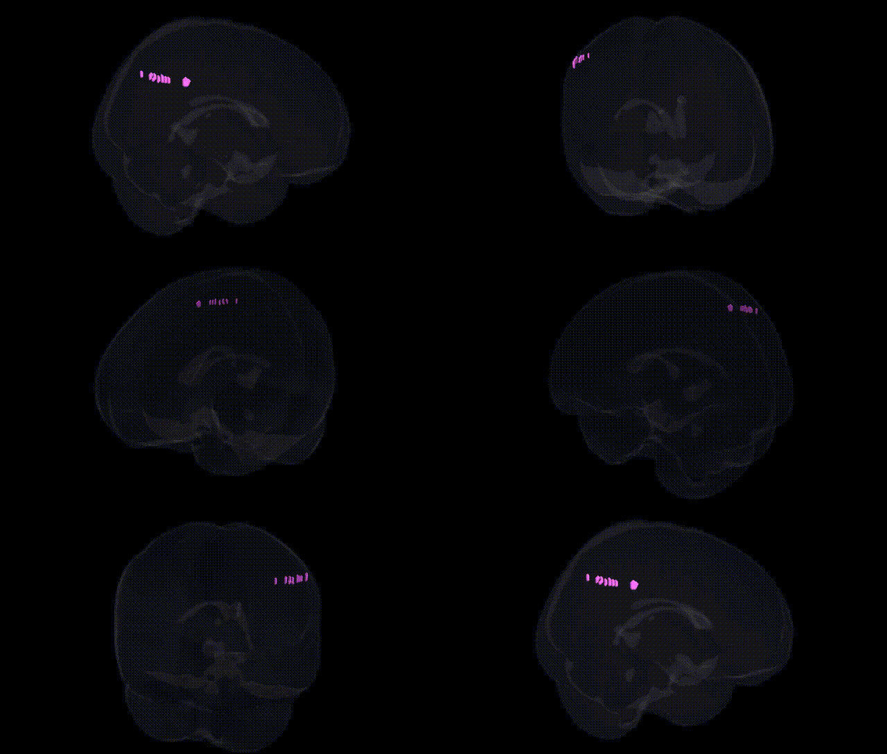
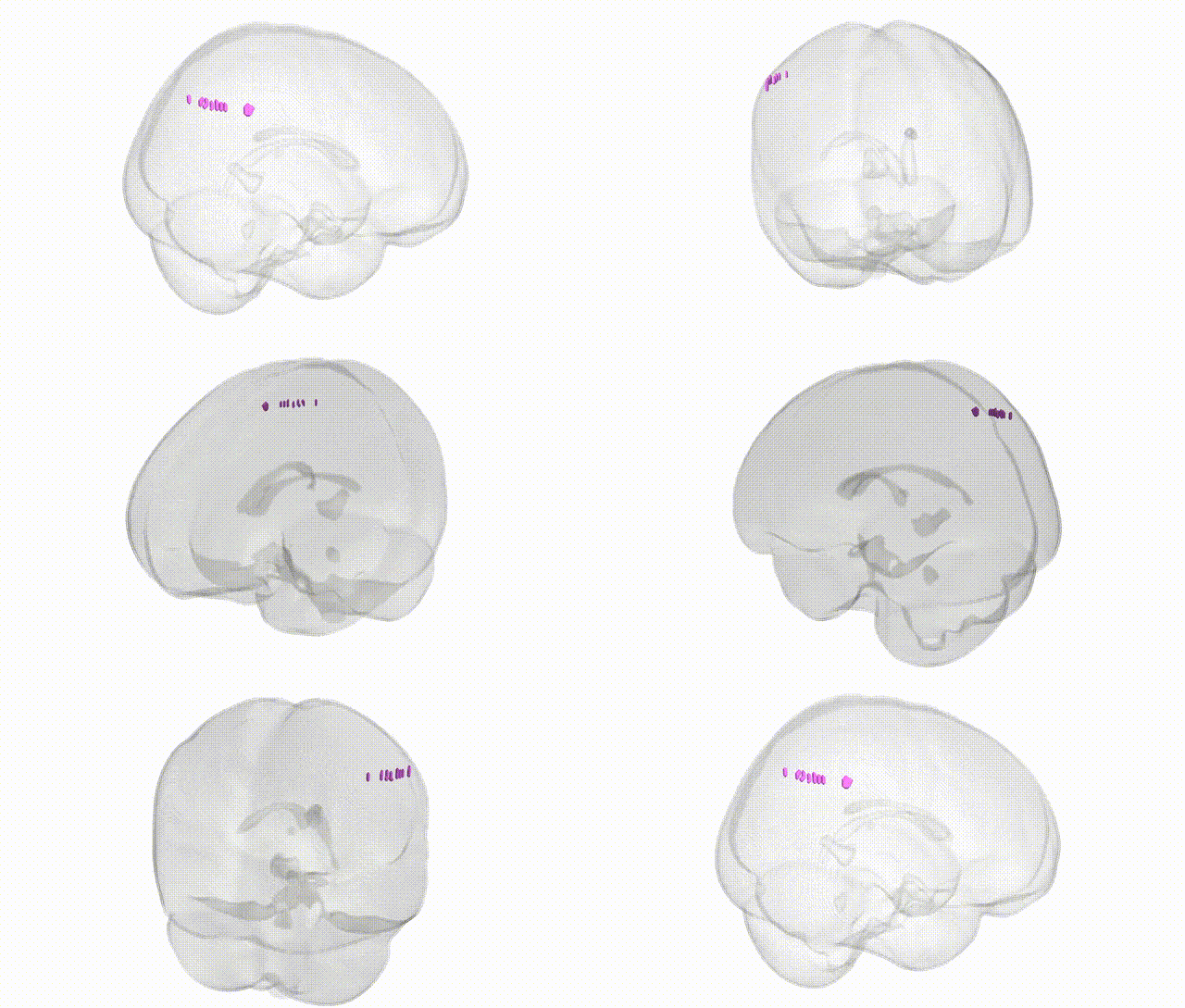
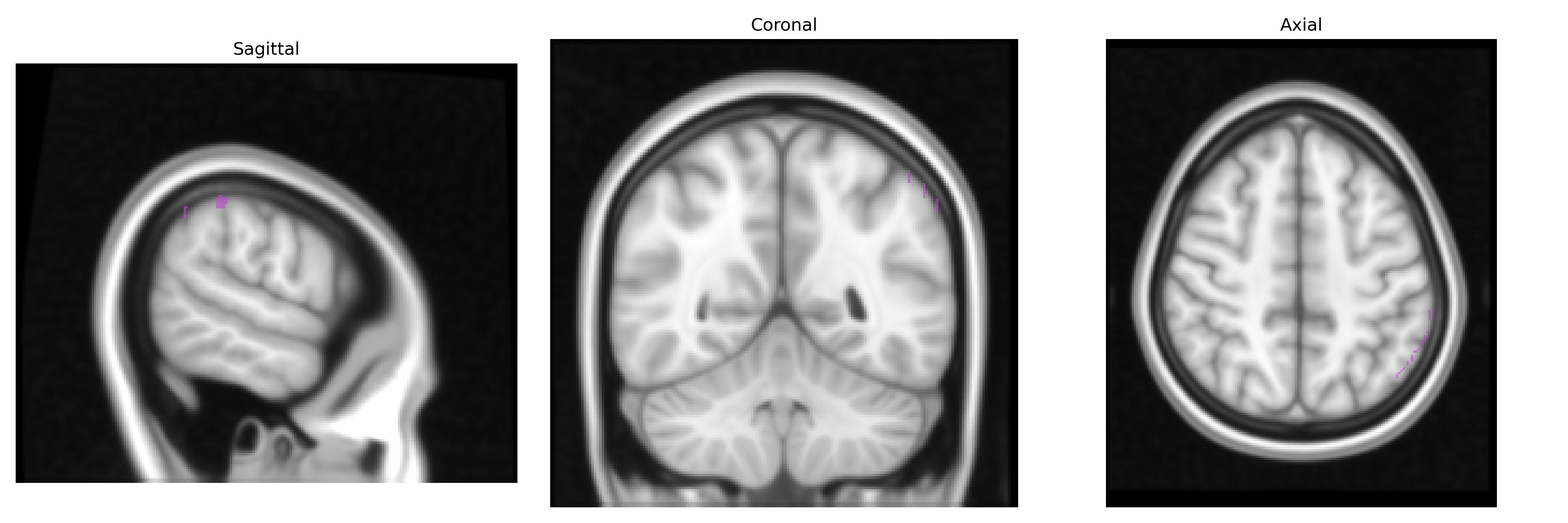
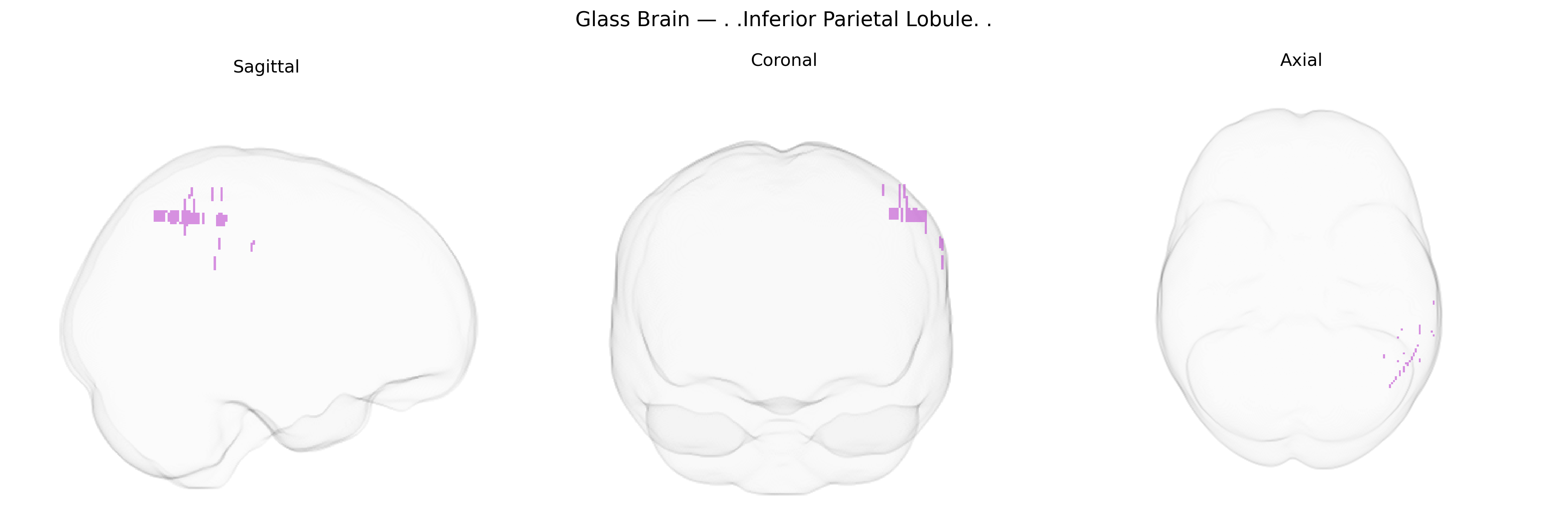
The bilateral Inferior Parietal Lobule (IPL) is a heteromodal association cortex located in the posterior parietal lobe, bordering the superior temporal and occipital regions, and typically comprising the supramarginal and angular gyri. It receives convergent visual, auditory, and somatosensory input and is critically involved in multimodal integration, spatial attention, body schema, language processing (including semantic and phonological aspects), number processing, and aspects of social cognition such as theory of mind and imitation. The IPL is functionally lateralized, with the left IPL more strongly associated with language and praxis, and the right IPL more prominent in visuospatial attention and neglect syndromes. Neuroimaging and lesion studies implicate this region in networks subserving attention reorienting, working memory, and the integration of sensory information into higher-order representations. Inferior parietal lobule
The bilateral inferior parietal lobule (IPL), as defined in the Talairach 1 mm atlas, has been implicated in a range of genetically influenced traits and disorders, primarily through imaging genetics and large-scale GWAS of brain structure and cognition. Variants in genes involved in synaptic plasticity, neurodevelopment, and connectivity—such as those in the BDNF, CNTNAP2, DISC1, and APOE loci—have been associated with IPL volume, cortical thickness, or functional activation, often in the context of working memory, language, and attentional control. Polygenic risk scores for schizophrenia, major depressive disorder, bipolar disorder, and autism spectrum disorder show convergent effects on IPL morphology and connectivity, consistent with the region’s role in default mode and frontoparietal networks. GWAS of cortical structure (e.g., ENIGMA and UK Biobank–based studies) have identified multiple loci with small effects on parietal morphology, including the inferior parietal cortex, linking genes involved in neuronal differentiation, axon guidance, and myelination to interindividual variation in this region. Additionally, genetic variants associated with Alzheimer’s disease risk (including APOE ε4 and loci such as CLU and PICALM) are related to altered IPL metabolism and atrophy patterns, while GWAS of cognitive abilities, educational attainment, and reading or mathematical skills highlight IPL-related circuits as key anatomical intermediaries, though individual variant effects on this specific Talairach-defined region remain modest and highly polygenic.
Overview generated by GPT-4o (2026).
Region ID: 933
Hemisphere: bilateral
Atlas: Talairach labels 1mm
Full Quality Version: Download MP4
Full Quality Version: Download MP4
Wali Sidiqyar*, Gaurav Rudravaram*, Elyssa M. McMaster, Trent M. Schwartz, Adam M. Saunders, Kurt G. Schilling, Bennett A. Landman "Introducing SPINS: A Shared Public Visualization Library of Neuroanatomical Structures." Medical Imaging with Deep Learning- short paper
This resource is licensed under CC0 1.0 Universal (Public Domain).