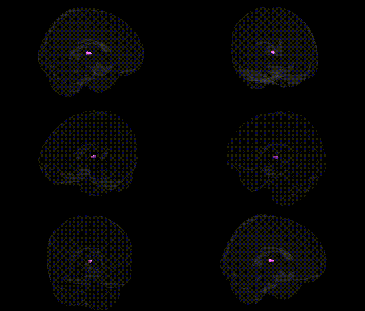
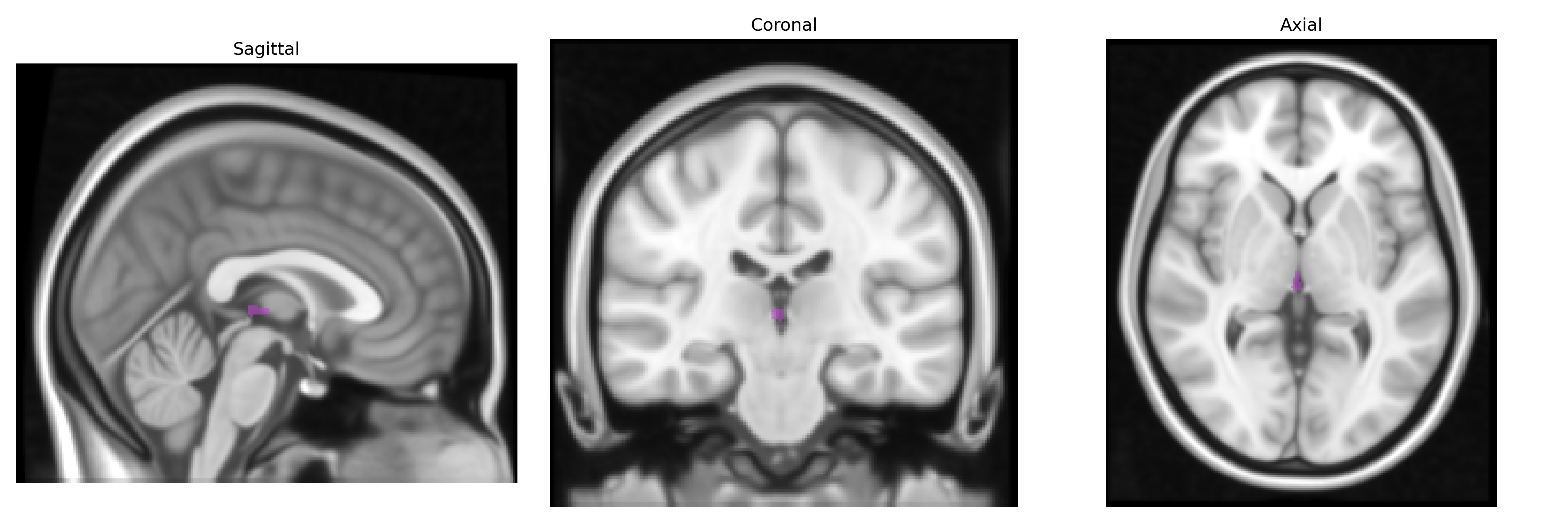
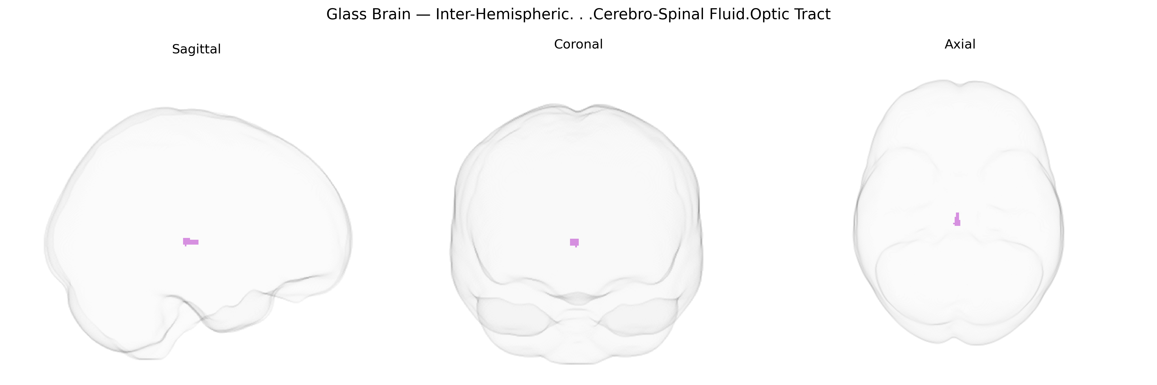
The bilateral inter-hemispheric cerebrospinal fluid (CSF) optic tract label in the Talairach 1 mm atlas likely corresponds to CSF-filled spaces and adjacent tissue along the midline near the optic chiasm and optic tracts, rather than a discrete parenchymal nucleus. Anatomically, this region encompasses the interhemispheric fissure and CSF compartments around the diencephalon and optic pathways, including cisterns and sulci where CSF circulates between the hemispheres and around the optic chiasm and tracts. Functionally, CSF in this area contributes to mechanical protection, buoyancy, and metabolic clearance for midline and visual pathway structures, while the nearby optic tracts relay visual information from the optic chiasm to the lateral geniculate nucleus and other visual targets. There is no direct Wikipedia article for this composite atlas label; a closely related structure is the Optic tract.
The bilateral inter-hemispheric cerebrospinal fluid (CSF) region near the optic tract, as defined in the Talairach 1 mm atlas, does not itself constitute a gray-matter structure with direct gene-expression or receptor-mapping data, and current genome-wide association studies (GWAS) rarely target CSF or atlas-defined CSF parcels as primary phenotypes. However, genetic studies of periventricular and interhemispheric CSF volumes, ventricular size, and related neuroimaging traits have implicated common variants in genes involved in neurodevelopment, brain morphogenesis, and fluid homeostasis—such as those in and around HMGA2, MAPT, and loci on chromosomes 3q28, 6q22, and 17q21—through large-scale imaging-genetics consortia (e.g., ENIGMA, UK Biobank). These loci are often pleiotropic, overlapping with GWAS signals for intracranial volume, white-matter integrity, and risk for neuropsychiatric or neurodegenerative disorders, including schizophrenia, Alzheimer’s disease, and other dementias, in which enlarged CSF spaces and altered periventricular morphology are common imaging markers. The optic tract itself, while not typically isolated in GWAS, lies along pathways implicated by genetic associations with visual pathway development and demyelinating conditions (e.g., multiple sclerosis), where variants in HLA regions and genes related to myelination and axonal integrity show robust effects, but the evidence links these genes to broader visual and white-matter network changes rather than specifically to the “inter-hemispheric CSF optic tract” label used in the Talairach atlas.
Overview generated by GPT-4o (2026).
Region ID: 624
Hemisphere: bilateral
Atlas: Talairach labels 1mm
Full Quality Version: Download MP4
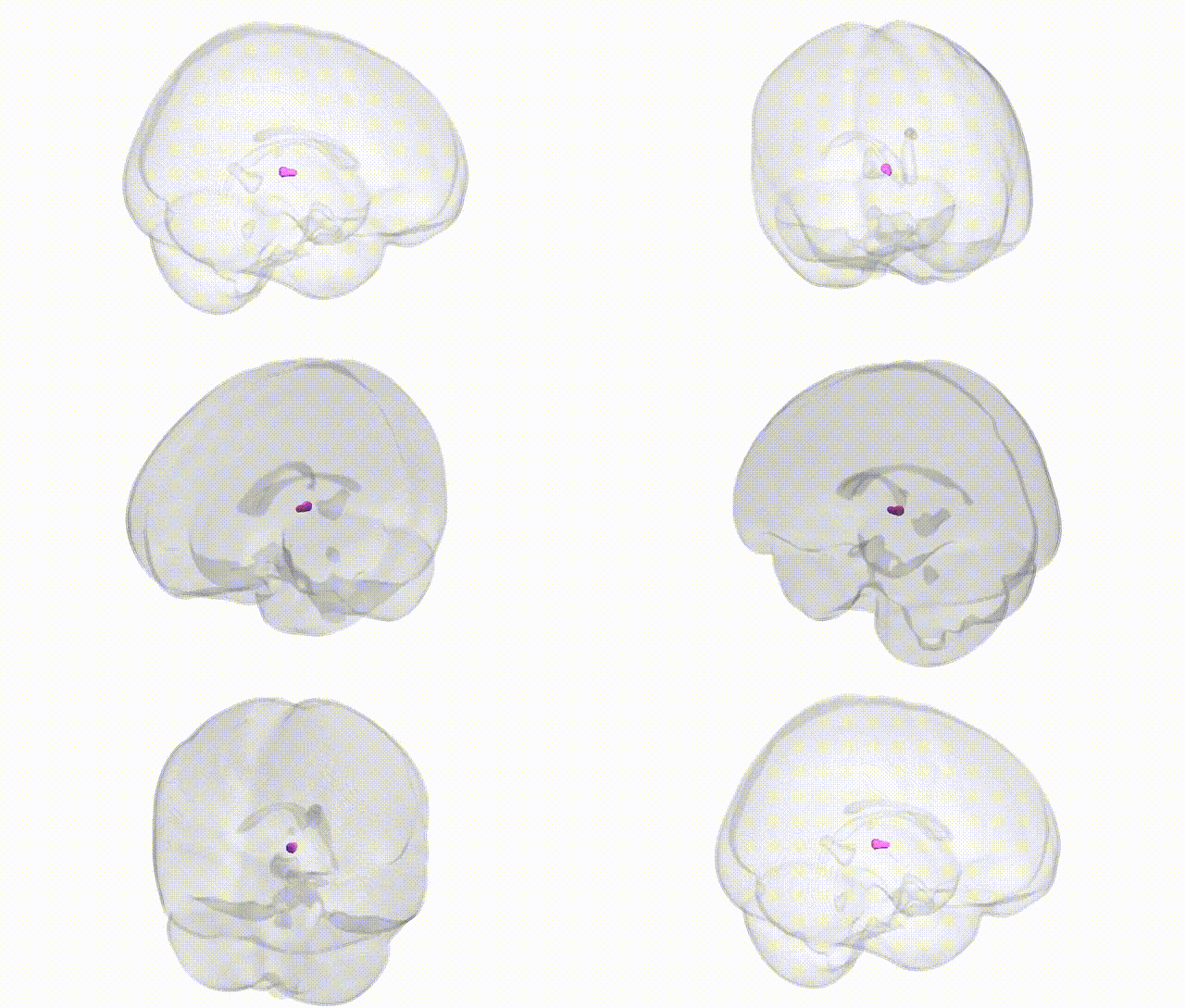
Full Quality Version: Download MP4
Wali Sidiqyar*, Gaurav Rudravaram*, Elyssa M. McMaster, Trent M. Schwartz, Adam M. Saunders, Kurt G. Schilling, Bennett A. Landman "Introducing SPINS: A Shared Public Visualization Library of Neuroanatomical Structures." Medical Imaging with Deep Learning- short paper
This resource is licensed under CC0 1.0 Universal (Public Domain).