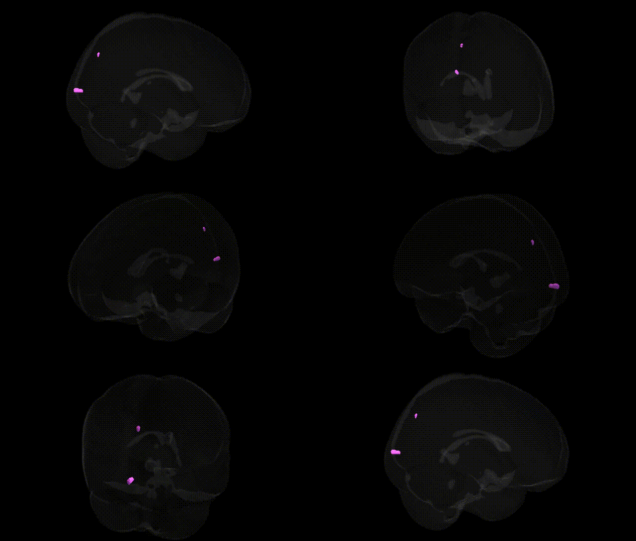
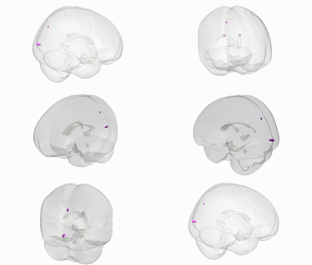
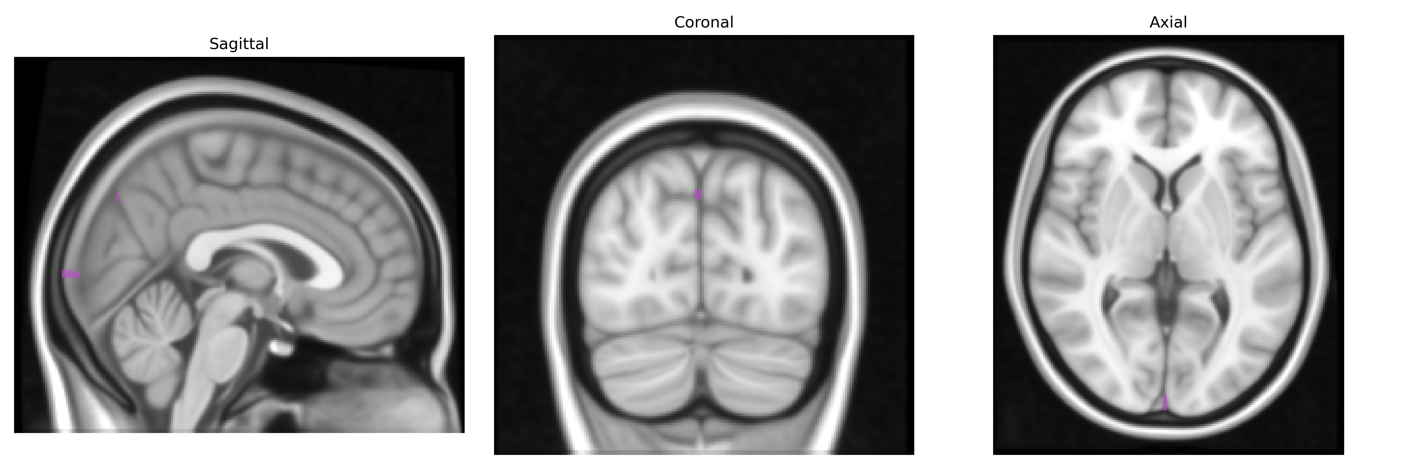
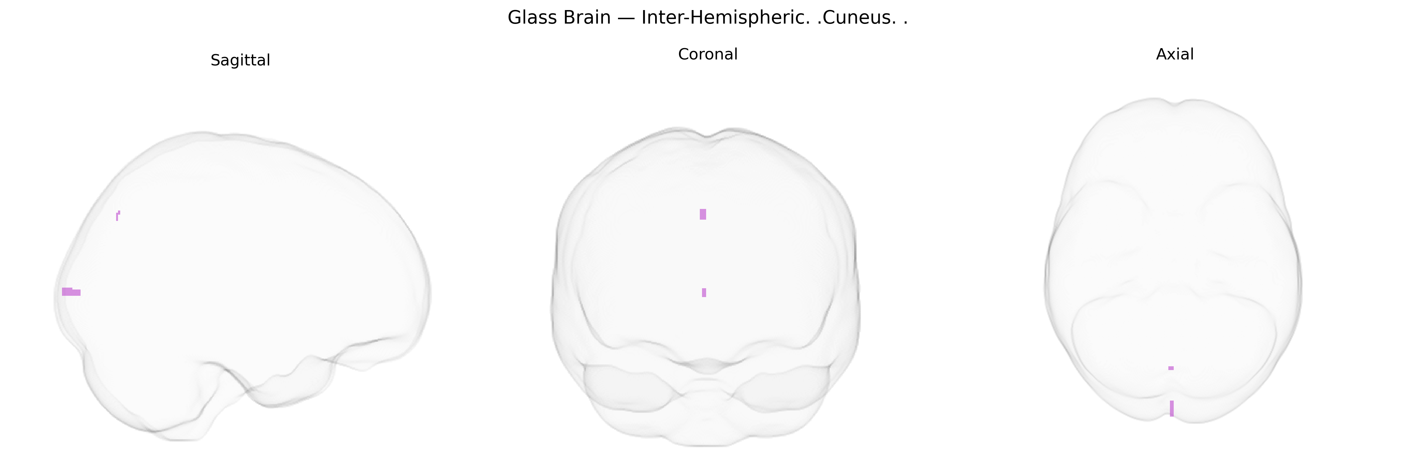
The bilateral inter-hemispheric cuneus refers to the medial occipital cortical region located in both hemispheres along the medial wall of the occipital lobe, bordering the longitudinal fissure. In the Talairach 1 mm atlas, this region encompasses gray matter dorsal to the calcarine sulcus and posterior to the parieto‑occipital sulcus, corresponding primarily to portions of the primary visual cortex (V1, Brodmann area 17) and adjacent visual association cortices (areas 18–19). It plays a key role in early visual processing, particularly of central visual fields, and participates in integration of visual information for perception of form, motion, and spatial relationships. As an inter-hemispheric label, it includes homologous cuneus tissue from both left and right hemispheres across the midline. Cuneus
The bilateral inter-hemispheric cuneus, a primary medial occipital region involved in early visual processing and higher-order visuospatial and attentional functions, has been implicated in multiple genetic imaging and GWAS-based endophenotype studies, though typically under broader “occipital” or “visual cortex” labels rather than specifically “Talairach 1 mm cuneus.” Twin and SNP-heritability work shows moderate to high heritability for cuneus volume, cortical thickness, and surface area, often linked to common variants in neurodevelopmental and synaptic genes (for example, MAPT region haplotypes, genes in Wnt signaling, and cell-adhesion pathways), and more generally to polygenic scores for global brain size and intracranial volume. Large-scale ENIGMA and UK Biobank imaging genetics analyses have associated cuneus morphology or functional activity with polygenic risk for schizophrenia, major depressive disorder, bipolar disorder, and autism spectrum disorder, where risk alleles in synaptic, glutamatergic, and calcium-channel genes (such as CACNA1C and related loci) show downstream associations with occipital and cuneus structure or activation patterns. GWAS of visual and cognitive traits, including visual acuity, reading and dyslexia-related measures, and general cognitive performance, report enrichment of visual cortex–expressed genes and link cuneus-related activation to variants in genes affecting myelination and axon guidance. Additional associations arise in GWAS of migraine, where variants in glutamatergic and cortical excitability pathways show effects on occipital/cuneus function, and in Alzheimer’s disease and related dementia risk loci, which have been tied via imaging-genetic mediation analyses to early posterior cortical atrophy patterns that prominently involve the cuneus and adjacent medial occipital cortex.
Overview generated by GPT-4o (2026).
Region ID: 538
Hemisphere: bilateral
Atlas: Talairach labels 1mm
Full Quality Version: Download MP4
Full Quality Version: Download MP4
Wali Sidiqyar*, Gaurav Rudravaram*, Elyssa M. McMaster, Trent M. Schwartz, Adam M. Saunders, Kurt G. Schilling, Bennett A. Landman "Introducing SPINS: A Shared Public Visualization Library of Neuroanatomical Structures." Medical Imaging with Deep Learning- short paper
This resource is licensed under CC0 1.0 Universal (Public Domain).