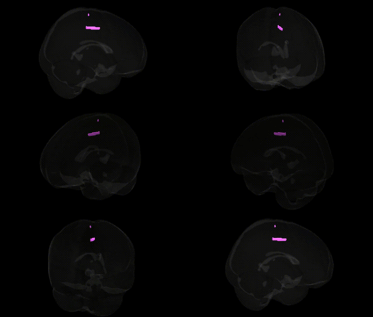
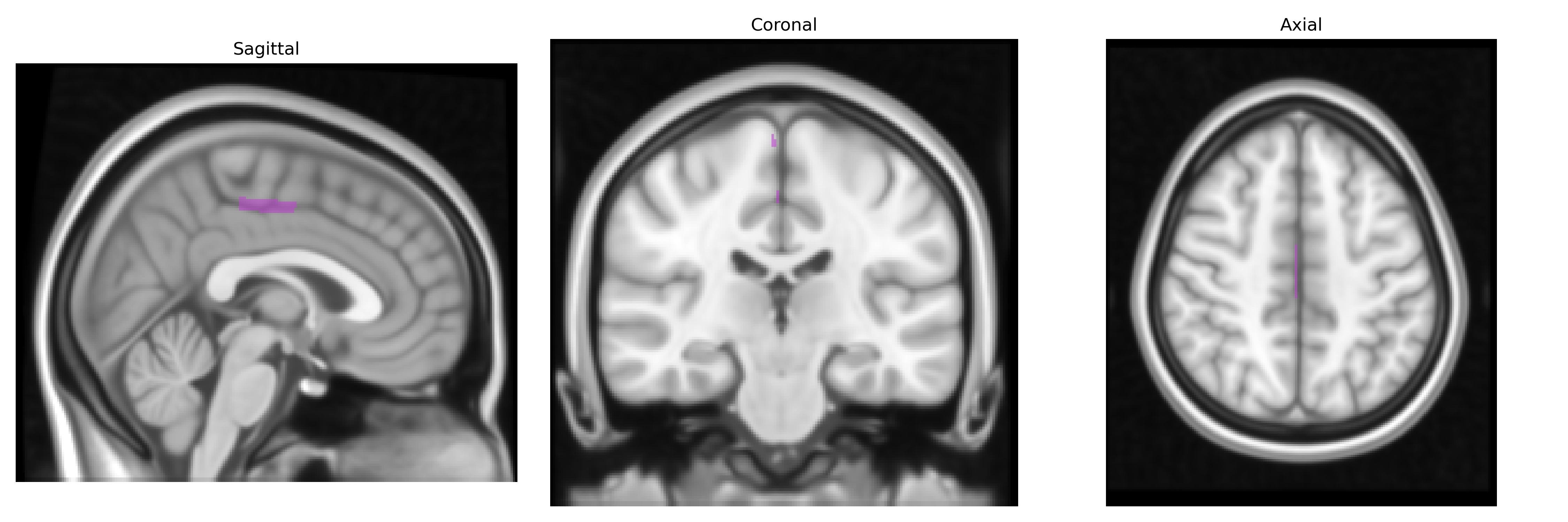
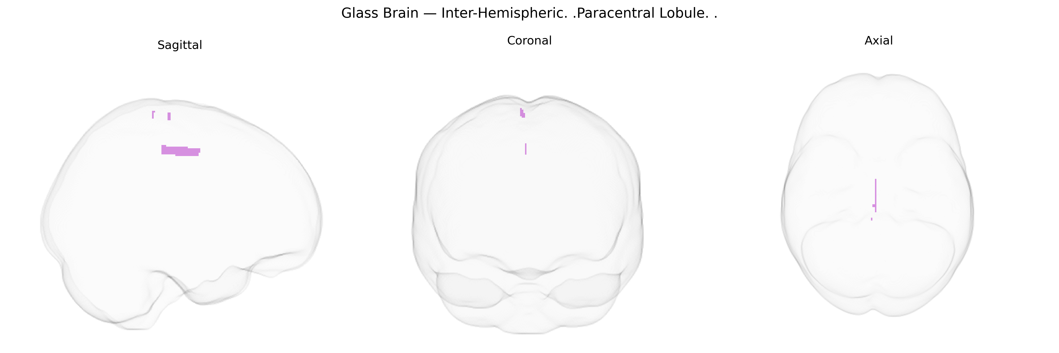
The bilateral inter-hemispheric paracentral lobule is a medial cortical region located on the superomedial surface of the cerebral hemispheres, bordering the central sulcus and encompassing portions of the primary motor and primary somatosensory cortices that represent the contralateral lower limb and perineal areas. Functionally, it participates in voluntary motor control and somatosensory processing of the leg and foot, contributes to sphincter control and aspects of pelvic floor function, and integrates sensorimotor information relevant to posture and gait. Its position along the midline inter-hemispheric fissure makes it a key hub for coordination of bilateral lower-extremity movements and for maintaining postural stability. There is no direct Wikipedia article for this exact Talairach label; see the related region: Paracentral lobule.
The bilateral inter-hemispheric paracentral lobule, encompassing medial primary motor and somatosensory areas for lower limbs, has been implicated in genetic studies primarily through large-scale GWAS of brain structure, motor function, and neuropsychiatric risk, although direct, region-specific associations are limited and often reported at coarser parcellations (e.g., medial sensorimotor or superior paracentral regions rather than the precise Talairach label). Imaging-genetics consortia such as ENIGMA and UK Biobank–based studies have identified heritable variation in cortical thickness and surface area of medial sensorimotor/paracentral regions associated with common variants in genes involved in neurodevelopment, synaptic function, and axon guidance (e.g., variants near MAPT, MIR137, and other neurodevelopmental loci), with overlaps across motor, cognitive, and psychiatric phenotypes. GWAS of lower-limb motor function, gait, and balance, as well as structural measures of the central sulcus and adjacent paracentral cortex, point to polygenic influences involving genes regulating corticospinal tract development and myelination, though specific loci are often broadly assigned to motor cortex rather than strictly to the paracentral lobule. Paracentral lobule involvement has also been reported in imaging-genetic studies of multiple sclerosis, amyotrophic lateral sclerosis, and neurodegenerative and neurodevelopmental disorders, where risk variants in immune-related and axonal integrity genes (e.g., HLA region, oligodendrocyte-related genes) correlate with atrophy or altered functional connectivity in medial sensorimotor regions. Overall, current evidence supports moderate heritability and polygenic influences on the structure and function of the paracentral lobule, with genetic associations typically emerging in the context of broader motor cortex, neurodegenerative, or psychiatric GWAS rather than as highly specific, paracentral-lobule–only findings.
Overview generated by GPT-4o (2026).
Region ID: 1047
Hemisphere: bilateral
Atlas: Talairach labels 1mm
Full Quality Version: Download MP4
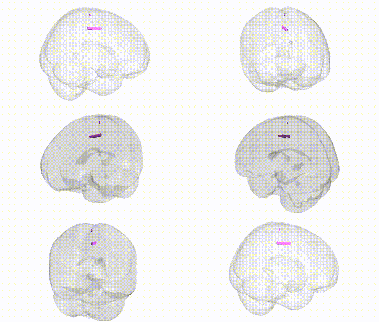
Full Quality Version: Download MP4
Wali Sidiqyar*, Gaurav Rudravaram*, Elyssa M. McMaster, Trent M. Schwartz, Adam M. Saunders, Kurt G. Schilling, Bennett A. Landman "Introducing SPINS: A Shared Public Visualization Library of Neuroanatomical Structures." Medical Imaging with Deep Learning- short paper
This resource is licensed under CC0 1.0 Universal (Public Domain).