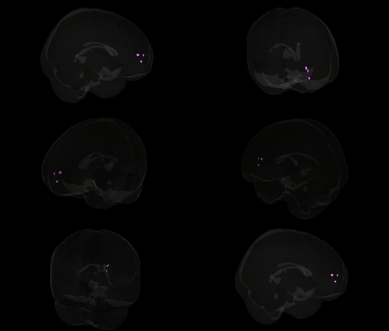
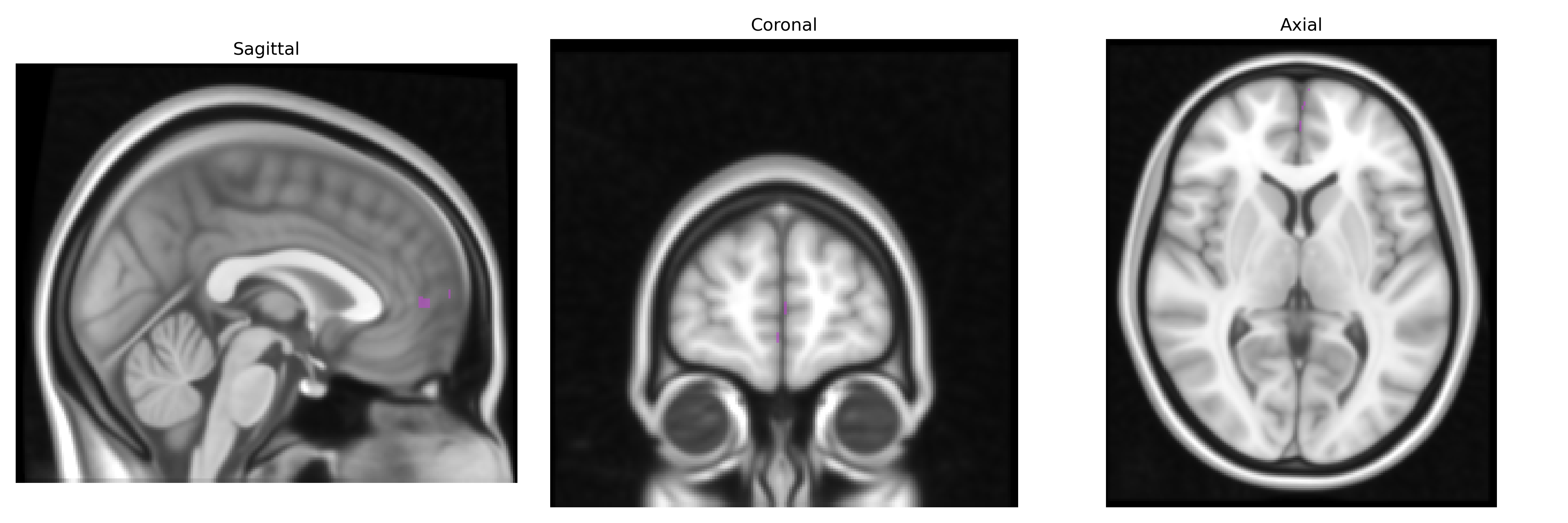
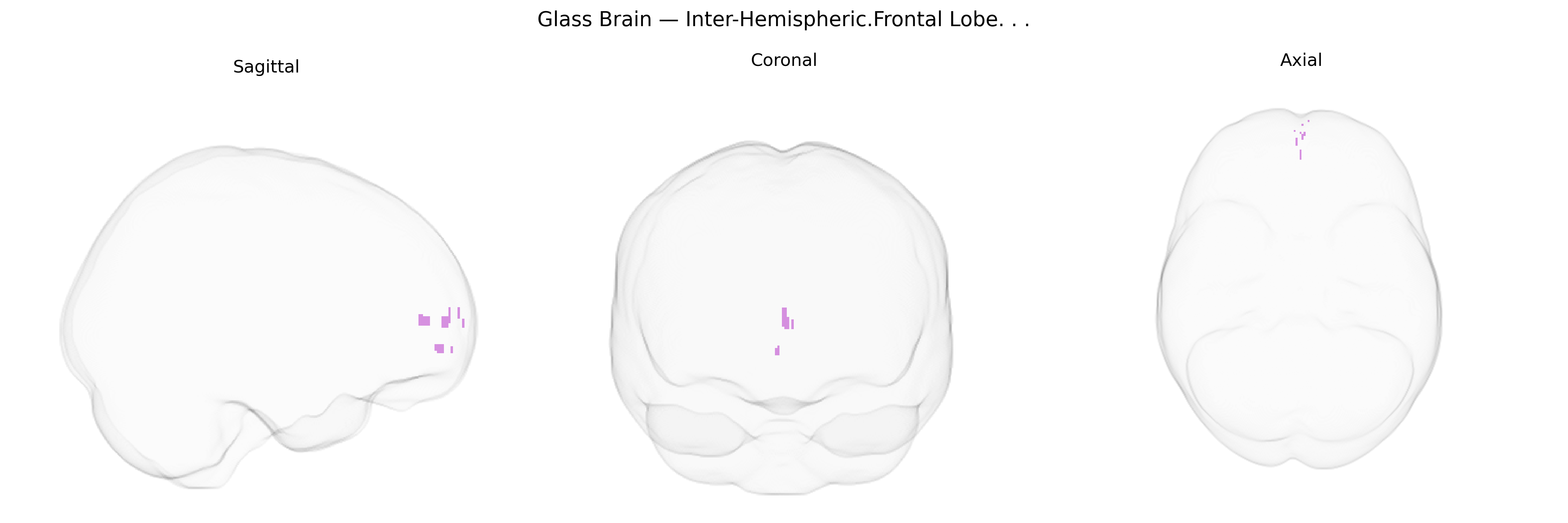
The bilateral Inter-Hemispheric.Frontal Lobe region in the Talairach 1 mm Atlas refers to medial frontal cortical territories located along and adjacent to the longitudinal fissure, encompassing interhemispheric aspects of the frontal lobes such as portions of the medial prefrontal cortex, supplementary motor area, and anterior cingulate-adjacent zones. Functionally, this interhemispheric frontal territory participates in higher-order cognitive control, including decision-making, performance monitoring, and aspects of working memory, as well as in motor planning and coordination of complex, often bilateral, actions. It is also implicated in social-cognitive processes and emotional regulation through its connections with limbic and subcortical structures. There is no direct Wikipedia article for “Inter-Hemispheric.Frontal Lobe”; a closely related structure is the Medial prefrontal cortex.
The bilateral inter-hemispheric frontal region, encompassing medial and superior frontal structures including parts of the medial prefrontal and anterior cingulate cortices, has been repeatedly implicated in genetic and genome-wide association studies through its role in higher-order cognition, emotion regulation, and psychopathology. Numerous GWAS of cortical thickness and surface area (e.g., ENIGMA, UK Biobank) have identified common variants in genes involved in neurodevelopmental and synaptic pathways—such as MAPT, GRIN2B, and genes in the Wnt and axon-guidance pathways—associated with structural variation in medial and superior frontal cortices, including inter-hemispheric frontal regions. Polygenic risk scores for schizophrenia, bipolar disorder, major depressive disorder, and ADHD show robust associations with altered volume or connectivity in medial frontal circuits, consistent with these regions’ involvement in default mode and executive networks. Specific risk loci for psychiatric conditions (e.g., CACNA1C, ZNF804A, DRD2) and for neurodevelopmental and cognitive traits (e.g., educational attainment, intelligence, and autism spectrum conditions) have been linked to functional and structural changes in midline frontal regions, including altered inter-hemispheric connectivity across the frontal midline. Additionally, genetic variants influencing callosal development and white matter integrity (e.g., in ROBO and SLIT family genes and other axon guidance molecules) have been associated with individual differences in frontal inter-hemispheric connectivity, thereby linking this Talairach-defined inter-hemispheric frontal area to a broad polygenic architecture underlying cognition, personality, and vulnerability to psychiatric and some neurodegenerative disorders.
Overview generated by GPT-4o (2026).
Region ID: 619
Hemisphere: bilateral
Atlas: Talairach labels 1mm
Full Quality Version: Download MP4
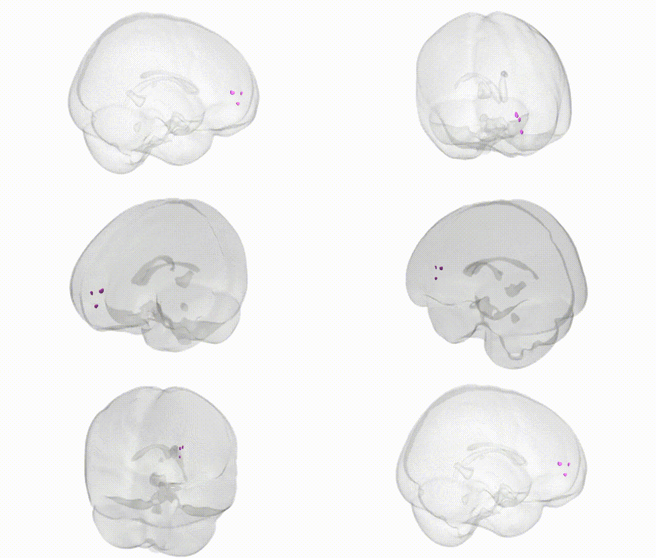
Full Quality Version: Download MP4
Wali Sidiqyar*, Gaurav Rudravaram*, Elyssa M. McMaster, Trent M. Schwartz, Adam M. Saunders, Kurt G. Schilling, Bennett A. Landman "Introducing SPINS: A Shared Public Visualization Library of Neuroanatomical Structures." Medical Imaging with Deep Learning- short paper
This resource is licensed under CC0 1.0 Universal (Public Domain).