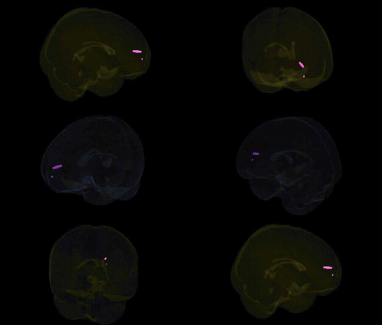
The bilateral Inter-Hemispheric Frontal Lobe Medial Frontal Gyrus corresponds primarily to the medial portion of the superior frontal gyrus along the interhemispheric fissure, encompassing parts of the medial prefrontal cortex and, more ventrally and posteriorly, regions that border on the anterior cingulate cortex. This region is involved in higher-order executive and cognitive control functions, including decision-making, performance monitoring, and aspects of working memory, as well as self-referential processing and components of the default mode network. Medially, it contains portions of Brodmann areas such as BA8, BA9, BA10, and BA32, depending on the exact Talairach coordinates, and participates in networks subserving attention, motivation, and the regulation of action. There is no single Wikipedia article dedicated specifically to the “medial frontal gyrus” as labeled in the Talairach 1 mm atlas; a closely related structure and functional counterpart is the Medial prefrontal cortex.
The medial frontal gyrus, a core component of the medial prefrontal cortex and anterior cingulate–adjacent networks, has been repeatedly implicated in genetic and GWAS-based imaging studies linking variation in this region to psychiatric, cognitive, and personality-related traits. Polygenic risk for schizophrenia, bipolar disorder, major depressive disorder, and autism spectrum disorder shows convergent effects on cortical thickness, surface area, or functional connectivity of medial frontal regions, including bilateral medial frontal gyrus, consistent with this area’s role in mood regulation, self-referential processing, and cognitive control. Large-scale ENIGMA and UK Biobank imaging-genetics consortia have identified common variants in genes involved in synaptic function and neurodevelopment (for example, CACNA1C, DRD2, GRIN2B, and several glutamatergic and GABAergic genes) that associate with medial frontal morphology or activity, often overlapping with loci conferring risk for psychiatric disorders. GWAS of resting-state networks and task-based activation have linked medial frontal gyrus connectivity to variants in genes such as ZNF804A and others influencing default mode and fronto-parietal networks, while studies of personality and cognitive traits (e.g., conscientiousness, neuroticism, intelligence, and executive function) have associated regional volume or activation in this region with polygenic scores for those traits. Additionally, structural and functional alterations of the medial frontal gyrus show heritability and are influenced by APOE and other Alzheimer’s disease–related loci, and have been observed in carriers of risk alleles for obsessive-compulsive disorder and attention-deficit/hyperactivity disorder, underscoring this region as a genetically sensitive hub for higher-order cognition and emotion.
Overview generated by GPT-4o (2026).
Region ID: 659
Hemisphere: bilateral
Atlas: Talairach labels 1mm
Full Quality Version: Download MP4
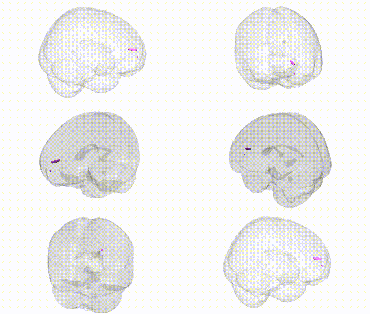
Full Quality Version: Download MP4
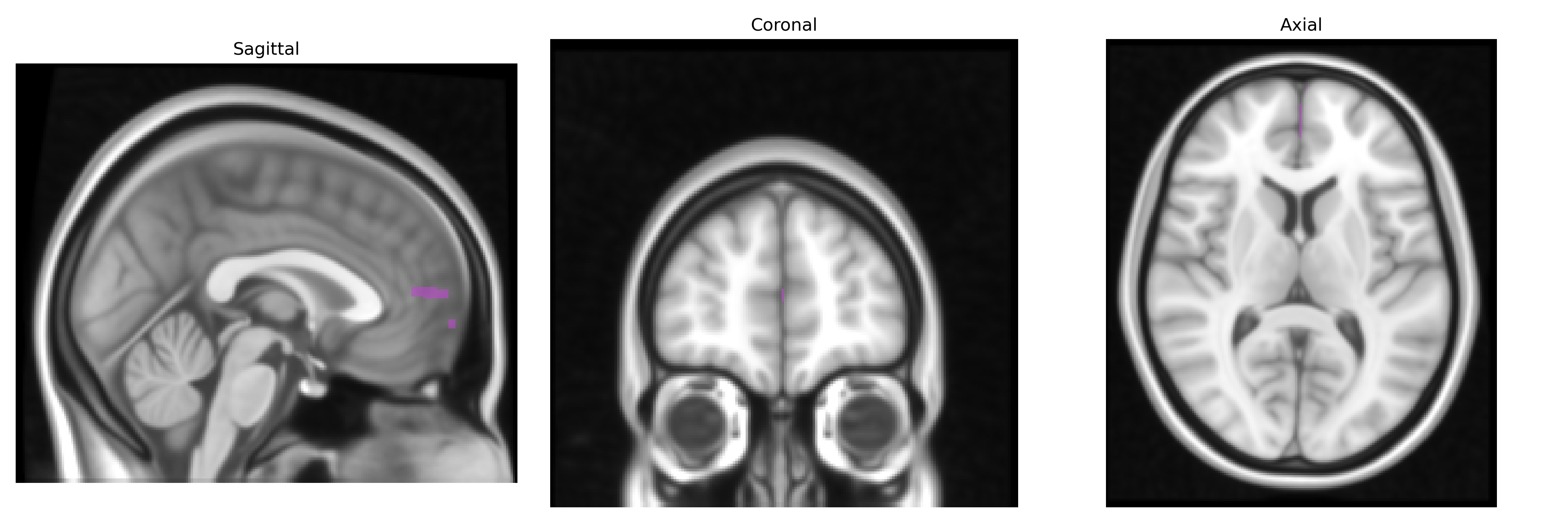
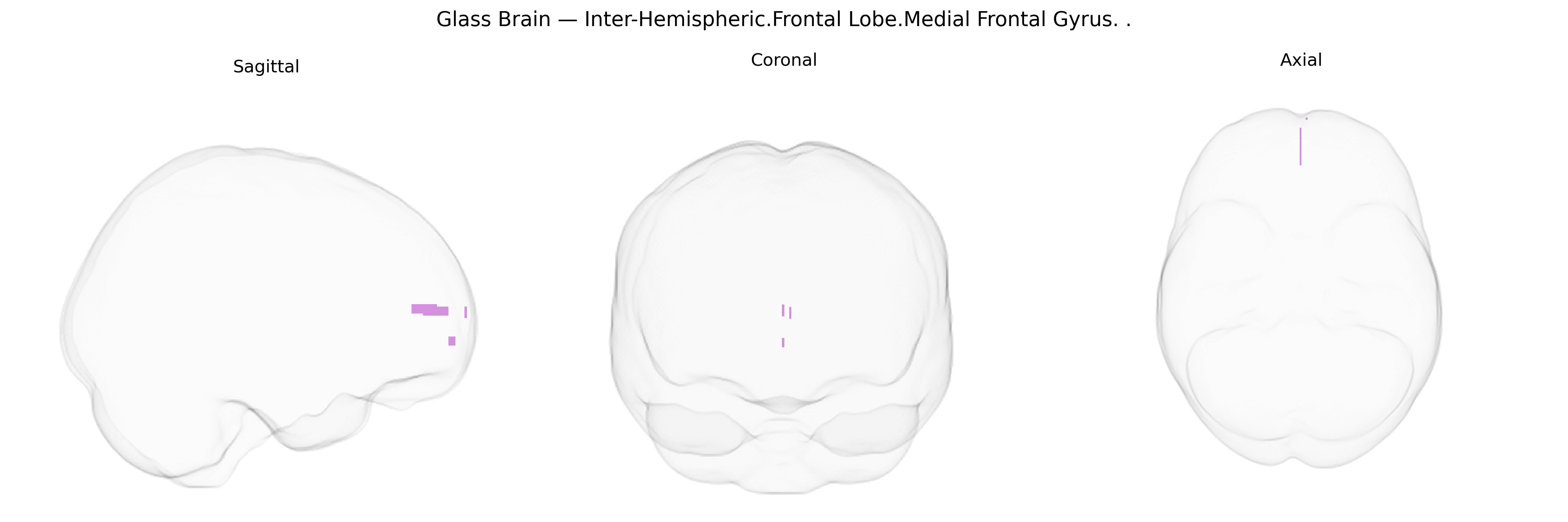
Wali Sidiqyar*, Gaurav Rudravaram*, Elyssa M. McMaster, Trent M. Schwartz, Adam M. Saunders, Kurt G. Schilling, Bennett A. Landman "Introducing SPINS: A Shared Public Visualization Library of Neuroanatomical Structures." Medical Imaging with Deep Learning- short paper
This resource is licensed under CC0 1.0 Universal (Public Domain).